


Services on Demand
Journal

Article
 text in
  English (pdf)
 Article in xml format
 Article references
 Send this article by e-mail
Indicators
-
 Cited by SciELO
Related links
-
 Similars in
SciELO
Similars in
SciELO
Share

 Permalink
Cirugía paraguaya
On-line version ISSN 2307-0420
Cir. parag. vol.47 no.3 Asunción Dec. 2023
https://doi.org/10.18004/sopaci.2023.diciembre.28
CASE REPORT
Choledocholithiasis resolved by laparoscopy. Case report
1
 http://orcid.org/0000-0002-2279-5620
1
http://orcid.org/0000-0003-0652-1985
1
http://orcid.org/0000-0002-7319-7479
1Instituto de Previsión Social, Hospital de Especialidades Quirúrgicas Ingavi. Asunción, Paraguay
Choledocholitiasis is a common obstructive pathology of the bile ducts, which can lead to a series of symptoms and complications. We present a 64-year-old female patient, with a history of cholecystectomy 30 years ago, who presented with epigastric pain, jaundice, choluria and acholia for six days. Upon admission obstructive jaundice related to gallstones was diagnosed by MRCP. The condition could not be resolved by ERCP and was resolved with a laparoscopic approach.
Keywords: choledocholithiasis; aparoscopic choledochotomy; ERCP
La coledocolitiasis es una patología obstructiva frecuente de las vías biliares, pudiendo llevar a una serie de síntomas y complicaciones. Presentamos una paciente femenina, de 64 años con antecedente de co- lecistectomía 30 años antes, que acude con dolor epigástrico de 6 días de evolución, ictericia, coluria y acolia. Al ingreso se diagnostica por colangioresonancia, ictericia obstructiva de etiología litiásica. El cuadro no pudo resolverse por colangiopancreatografía retrógrada endoscópica y se resuelve por vía laparoscópica.
Palabras clave: coledocolitiasis; coledocotomia videolaparoscópica; CPRE
INTRODUCTION
Choledocholitiasis is one of the most frequent pathologies of the bile ducts and consists in the presence of calculi on the common bile duct, which obstructs bile flow causing complications such as: obstructive jaundice, acute pancreatitis, acute cholangitis, among others 1. Choledocholitiasis can be classified in primary and secondary, with primary being the ones that develop within 24 months of the previous cholecystectomy in absence of obstructive symptoms, while secondary being the ones that develop within 24 months of the previous bile surgery. For the diagnosis it is necessary to perform a proper anamnesis, laboratorial diagnosis of the jaundice and ob- structive pattern, besides utilizing complementary studies and images, being magnetic resonance cholangiopancreatography (MRCP) the gold standard for the diagnosis of choledocholitiasis 2. The therapeutic gold standard is the endoscopic retrograde cholangiopancreatography (ERCP) which allows the ex- traction of these type of calculi, and if the patient presents very strong criteria, it could be performed even without a previous MRCP, being these the criteria: evidence of choledocholitiasis by ultrasonography, total bilirubin over 4 mg/dl, or signs and symptoms of ascending cholangitis. The ERCP, besides being a therapeutic method it could also be utilized as a diagnosis method, considering the 95% sensibility and specification for the detection of calculi on the bile ducts. 2,3. There are situations when the extraction of calculi cannot be solved by endoscopic approach: when calculi of considerable size are present, disproportionate to the common bile duct’s diameter, or when identification of the duodenal papilla is unsuccessful. When the ERCP is not possible, the surgical resolution must be pro- posed, with the laparoscopic approach being the one chosen, along with choledochotomy and calculi extraction, and placement of a T-tube for the bile duct’s decompression.
CLINICAL CASE
A 64-year-old female patient presented with a 6-day history of epigastric pain of insidious onset, colic type of moderate intensity, radiating to the interscapular zone and partially yields with common analgesic intake. Two days before admission, nausea and vomiting of food content on two occasions were added to the symptoms, and 24-hours before admission, change in skin and mucous membrane coloration which gain a yellowish tone were also added, besides choluria and acholia. For patholog- ical history, the patient reported arterial hypertension in regular treatment with 50 mg/day of losartan and denies other base pathologies. For surgical history, she reported a choledochotomy performed 30 years ago by Kocher incision. The ultrasound showed intrahepatic non-dilated bile ducts, and a 10.2mm common bile duct on its proximal portion, with the distal portion not being visible. On admission, the laboratory reported: white blood cells 17,600/µL, neutrophils 76%, hemo- globin 12.3 g/dL, total bilirubin 2.66 mg/dL and direct bilirubin
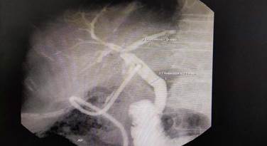
1.41 mg/dL, alkaline phosphatase 171 U/L, GOT 51 U/L and GPT 71 U/L. Due to laboratory findings, the patient was sub- mitted to a MRCP which showed the common hepatic duct and common bile duct to up to 10 mm, with a 5 mm signal voids compatible with calculi. An ERCP was performed based on the aforementioned findings, in which the duodenal papilla could not be identified, therefore, the extraction of choledocholithiasis was not possible and so surgical therapy was performed: laparoscopic choledochotomy with calculi extraction with a Fogarty balloon, peroperative cholangiography and T-tube placement (see Figure 1). In the peroperative cholangiography, proper contrast passage to the duodenum without residual filling defect’s imagery was recorded. The patient had a favorable postoperative course, beginning oral tolerance within the first postoperative day (POD1). Within the POD5 a T-tube cholan- giogram was performed, in which proper contrast passage to the duodenum was recorded, without evidence of leaking or signs of signal voids suggesting residual calculi (see Figure 2). The patient is discharged on the POD6 without complications, good oral tolerance, no spontaneous pain nor infection and it is planned for the T-tube to be extracted within POD45. The extraction of the T-tube was performed after the POD45 and annual controls are performed on the patient without complications up to this date.
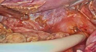
Figure 1. Entrance of the T-tube to the place where the choledochotomy was perfor- med is observed, along with the suture points.
DISCUSSION
The presence of choledocholithiasis as a bile pathology is very frequent on our region, with the ERCP being the chosen method upon its presence for the calculi extraction and the prophylactic cholecystectomy within 48-hours after the performed ERCP to avoid future complications. Surgical resolution must proceed when the choledocholithiasis cannot be resolved by an endo- scopic approach4, with the choices being the open or laparoscop- ic approach, given that a gold standard for surgical treatment does not exist. Several studies, both regional and international, conclude that choledochotomy and extraction of calculus with or without placement of a T-tube for draining is a safe option of treatment2,4-7 and according to the findings of Yegros-Ortiz et al7 there is no significant difference in the morbi-mortality with or without the placement of the T-tube after the choledochotomy. Canullan et al2 proposes the efficacy superiority of the transcystic approach of extraction over the transcholedocian approach.
In our case the choledochotomy with T-tube placement was decided due to the absence of a previous endoscopic papillotomy, which implicated the placement of a drainage for bile duct de- compression.
REFERENCES
1. Barreras González JE, Ruiz Torres J, Torres Peña R, Martínez Alfonso MA, Faife Faife BC, Hernández Gutiérrez JM, et al. Coledocolitiasis: Opciones actuales de tratamiento laparoscópico y endoscópico. Rev haban cienc méd [Internet]. 2010 Sep [citado 2023 Oct 15];9(3): 374-384. Disponible en: Disponible en: http://scielo.sld.cu/scielo.php?script=sci_arttext&pid=S1729-519X2010000300012&lng=es . [ Links ]
2. Canullán CM, Petracchi EJ, Baglietto N, Zandalazini HI, Quesada BM, Merchán del Hierro P, et al. Abordaje laparoscópico sistemático de la coledocolitiasis. Rev. argent. cir. [Internet]. 2021 Abr [citado 2023 Oct 15];113(1):62-72. Disponible en: Disponible en: http://dx.doi.org/10.25132/raac.v113. n1.1501.ei . [ Links ]
3. LaArtifon E, Tchekmedyian AJ, Aguirre PA. Colangiopancreatrografía retrógrada endoscópica: una técnica en permanente evolución. Rev. gastroenterol. Perú [Internet]. 2013 Oct [citado 2023 Oct 15];33(4):321-327. Disponible en: Disponible en: http://www.scielo.org.pe/scielo.php?script=sci_ arttext&pid=S1022-51292013000400006&lng=es . [ Links ]
4. Manterola C, Pineda V, Vial M. Efectividad del tratamiento laparoscópico de la colelitiasis y la coledocolitiasis: Revisión global de la evidencia. Rev Chil Cir [Internet]. 2007 Jun [citado 2023 Oct 15];59(3):198-207. Disponible en: Disponible en: http://www.scielo.cl/scielo.php?script=sci_arttext&pid=S0718-40262007000300006&lng=es . Disponible en: http://www.scielo.cl/scielo.php?script=sci_arttext&pid=S0718-40262007000300006&lng=es. http://dx.doi.org/10.4067/S0718-40262007000300006 . [ Links ]
5. Álvarez-Chica LF, Rico-Juri JM, Carrero-Rivera SA, Castro-Villegas F. Coledocolitiasis y exploración laparoscópica de la vía biliar. Un estudio de cohorte. Rev. Colomb. cir. [Internet]. 2021 June [citado 2023 Oct 15];36(2):301-311. Disponible en: Disponible en: http://www.scielo.org.co/scielo.php?script=sci_arttext&pid=S2011-75822021000200301&lng=en [ Links ]
6. Ramírez P, Samaniego C, Ortiz Villalba JM. Litiasis de la vía biliar principal: resultados del tratamiento quirúrgico y de la papilotomía endoscópica. Rev. Cir. Parag. [Internet]. 2014 June [citado 2023 Oct 15];38(1):8-11. Disponible en: Disponible en: http://scielo.iics.una.py/scielo. php?script=sci_arttext&pid=S2307-04202014000100002&lng=en [ Links ]
7. Yegros-Ortiz CD, Acosta-López K, Montiel-Alfonso MA, Ferreira-Bogado M. Manejo videolaparoscópico de la coledocolitiasis en el servicio de cirugía general del Hospital Nacional - Itauguá. Cir. Parag. [Internet]. 2021 Aug [citado 2023 Oct 15];45(2):25-28. Disponible en: Disponible en: http://scielo.iics.una.py/scielo.php?script=sci_arttext&pid=S2307-04202021000200025&lng=en . [ Links ]
Author’s contributions: PESE, MFD, AJBM, GPVF and GDB contributed the idea, writing of the work, literature search and final approval.
Ethical considerations: During this work the ethical requirements towards the protection of the patient and their rights have been respected, as for the protection of privacy and assurance of free participation.
Received: January 08, 2023; Accepted: November 27, 2023
 Este es un artículo publicado en acceso abierto bajo una licencia Creative Commons
