




Serviços Personalizados
Journal

Artigo
 texto em
 Inglês (pdf)
 Artigo em XML
 Referências do artigo

 Tradução automática
 Enviar este artigo por email
Indicadores
-
 Citado por SciELO
Links relacionados
-
 Similares em
SciELO
Compartilhar

 Permalink

Cirugía paraguaya
versão On-line ISSN 2307-0420
Cir. parag. vol.49 no.1 Asunción abr. 2025
https://doi.org/10.18004/sopaci.2025.abril.24
REVIEW ARTICLE
Paraguayan Consensus regarding diagnosis and treatment of acute pancreatitis in adults. Guide for surgeons - 2025
1
2
 http://orcid.org/0000-0003-1988-7134
1
2
http://orcid.org/0009-0002-1294-5274
1
2
http://orcid.org/0000-0002-4121-3219
3
http://orcid.org/0000-0002-1696-8484
1
2
http://orcid.org/0000-0002-6194-7180
4
http://orcid.org/0000-0002-3693-9154
5
http://orcid.org/0000-0001-8952-6399
1
http://orcid.org/0000-0002-3316-2837
6
http://orcid.org/0000-0001-7417-4431
4
http://orcid.org/0009-0008-6314-7241
7
http://orcid.org/0009-0008-1401-336X
8
http://orcid.org/0000-0002-0365-1517
1
9
http://orcid.org/0000-0002-2945-0852
2
http://orcid.org/0009-0007-7439-4427
10
http://orcid.org/0009-0000-7058-3035
1
http://orcid.org/0000-0003-3255-5345
3
http://orcid.org/0009-0005-4214-6424
11
http://orcid.org/0009-0009-4721-7323
12
http://orcid.org/0009-0001-9790-2953
6
http://orcid.org/0000-0003-1245-2664
1
13
http://orcid.org/0000-0002-3545-5887
3
http://orcid.org/0000-0003-4286-3931
1Hospital de Clínicas. Asunción, Paraguay
2Hospital Central - Instituto de Previsión Social. Asunción, Paraguay
3Sanatorio Privado. Asunción, Paraguay
4Hospital San Jorge. Asunción, Paraguay
5Hospital Nacional de Itauguá. Itauguá, Paraguay
6Hospital Instituto de Previsión Social INGAVI. Fernando de la Mora, Paraguay
7Hospital San Ignacio - Misiones. Misiones, Paraguay
8Hospital de Ciudad del Este, Paraguay
9Hospital de Luque. Luque, Paraguay
10Hospital de Cnel. Oviedo. Coronel Oviedo, Paraguay
11Hospital de Concepción. Concepción, Paraguay
12Hospital de Filadelfia. Filadelfia, Paraguay
13Hospital Militar . Asunción, Paraguay
14Hospital de Encarnación. Encarnación, Paraguay
This document outlines the consensus reached by Paraguayan experts on the handling of acute pancreatitis (AP). The objective is to unify diagnostic and treatment criteria based on scientific evidence adapted to the local context, aiming to improve the quality of care and promote research in this field.
The methodology involved selecting 24 experts through a national survey, who were then organized into three working groups to address different aspects of AP. A comprehensive review of the scientific literature was conducted, covering publications from 1990 to 2024.
The document defines AP as an acute inflammatory process of the pancreas, with biliary lithiasis and alcohol consumption as the leading causes. Severity is classified as mild, moderate, severe, or critical, based on the presence of necrosis, organ failure, and complications. It highlights the importance of clinical parameters, laboratory tests (lipase and amylase), and imaging studies.
Use of the Petrov or revised Atlanta classification is recommended to assess the severity of AP, along with the Marshall scoring system to evaluate systemic complications.
CT scanning is considered essential for grading severity and diagnosing complications, particularly between the third and tenth day of disease progression. Ultrasound is emphasized as a valuable initial tool for identifying biliary causes and detecting early complications.
Ultimately, the goal is to standardize the handling of AP and improve patient outcomes with this pathology in Paraguay.
Keywords: acute pancreatitis; diagnostic; classification; treatment
Este documento detalla el consenso generado por expertos paraguayos en el manejo de la pancreatitis aguda (PA). El objetivo es unificar criterios de diagnóstico y tratamiento basados en la evidencia científica, adaptada al contexto local, buscando mejorar la calidad de la atención y promover la investigación en esta área. La metodología empleada incluyó la selección de 24 expertos a través de una encuesta nacional, quienes se organizaron en tres mesas de trabajo para abordar diferentes aspectos de la PA. Se realizó una revisión exhaustiva de la literatura científica en bases a publicaciones que datan de 1990 a 2024. El documento define la PA como un proceso inflamatorio agudo del páncreas, con la litiasis biliar y el alcohol como principales causas. La gravedad es clasificada en leve, moderada, grave y crítica, basándose en la presencia de necrosis, falla orgánica y complicaciones. Se destaca la importancia de los parámetros clínicos, laboratorio (lipasa y amilasa) y los estudios de imágenes. Se recomienda el uso de la clasificación de Petrov o Atlanta revisada para establecer la gravedad de la PA y la clasificación de Marshall para determinar las complicaciones sistémicas. La TAC es considerada fundamental para la clasificación de gravedad y el diagnóstico de complicaciones, especialmente entre el tercer y décimo día de evolución. Se enfatiza la utilidad de la ecografía como herramienta inicial para identificar la causa biliar y detectar complicaciones tempranas. Se busca - finalmente - estandarizar el manejo de la PA y mejorar los resultados en los pacientes con esta patología en el Paraguay.
Palabras claves: pancreatitis aguda; diagnóstico; clasificación; tratamiento
Special considerations
It should be noted that the diagnosis and management of patients with acute pancreatitis require a comprehensive approach that combines clinical, surgical, biochemical, and imaging evaluations. This combination represents the minimum requirement for the proper treatment of these patients.
Transfer patients with acute pancreatitis to a high-complexity hospital as soon as possible when:
- no clinical improvement is observed,
- abdominal pain increases,
- fever exceeds 38°C,
- white blood cell count, and CRP levels are rising, and/or
- imaging studies show findings compatible with local complications.
A high-complexity hospital is a referral medical center that provides highly specialized care. It is equipped with advanced technology (laboratory, ultrasound, CT scan, MRI, endoscopy, interventional procedures, operating rooms) and staffed by highly qualified professionals capable of managing complex diseases and critical conditions. It offers a wide range of medical and surgical specialties, as well as subspecialties such as neurosurgery, cardiovascular surgery, organ transplantation, advanced oncology, intensive care, endocrinology, infectious diseases, physiotherapy, among others. Additionally, it includes specialized areas such as intermediate and intensive care units, with these services operating 24 hours a day.
Introduction
Acute pancreatitis (AP) is an acute inflammatory condition of the pancreas, with a rising incidence. It presents across a broad clinical spectrum, ranging from mild, self-limiting forms to severe cases involving pancreatic necrosis and multiorgan dysfunction, which may lead to patient death.
The etiology of AP varies by geographic region, with biliary lithiasis and alcohol consumption being the most common causes worldwide. In Paraguay, biliary lithiasis remains the leading cause of AP, making timely diagnostic and therapeutic intervention essential for these patients.
Despite advances in the understanding of the pathophysiology of AP, as well as improvements in imaging techniques, intensive care, and interventional procedures, the severe form of the disease continues to show high mortality rates. This highlights the need to enhance strategies for the prevention, diagnosis, and treatment of this condition.
AP is a significant public health issue both globally and in Paraguay, with considerable social and economic impact on the population and healthcare systems. Addressing this problem requires a comprehensive approach that includes prevention, early diagnosis, appropriate treatment, and ongoing research.
The incidence of AP has increased in recent decades in the region, possibly due to lifestyle changes such as rising alcohol consumption and obesity.
Biliary lithiasis and alcohol consumption are the main causes in South America; similarly, the increase in blunt abdominal trauma from traffic accidents contributes to the condition.
All these factors lead to prolonged hospitalization, high treatment costs, and loss of productivity, negatively impacting the quality of life of patients and their families.
In Paraguay, biliary lithiasis is the leading cause of acute AP, highlighting the need to strengthen detection and treatment programs for this condition. Despite advances in the management of AP, the severe form of the disease continues to show a high rate of morbidity and mortality, underscoring the importance of improving diagnostic and therapeutic strategies.
It is essential to implement primary and secondary prevention strategies to reduce the incidence of AP, including awareness campaigns about the risks of excessive alcohol consumption, the need for treatment of gallbladder lithiasis, and the importance of maintaining a healthy weight.
Objectives
1. Standardize criteria and recommendations based on scientific evidence and adapted to the national context.
2. Improve the quality of care received by patients with acute AP in Paraguay.
3. Reduce mortality and complications through the identification of risk factors, promotion of early diagnosis, initial treatment, as well as the transfer of severe cases to other medical centers with intensive care units and access to appropriate procedures when necessary.
4. Promote research that enables the identification of areas where further investigation on AP is needed in Paraguay, such as the geographic distribution of the disease, risk factors within the local population, and the effectiveness of various prevention and treatment strategies.
5. Facilitate ongoing training for surgeons, gastroenterologists, and medical professionals in general.
Methodology
1-Expert selection and worktables organization: The experts were chosen through a nationwide anonymous virtual survey. Initially, 24 renowned expert surgeons from various institutions across the country were invited. The participants were selected based on their experience and knowledge in managing this condition.
3 worktables were organized: Section 1 (focused on general considerations, definitions, and diagnosis); Section 2 (on initial management); and Section 3 (on complications). Each group was composed of 8 experts coordinated by 2 members of the Executive Committee of the Paraguayan HPB Chapter.
2-Research and selection of scientific evidence: An exhaustive search of scientific information was conducted in the main biomedical databases: CENTRAL (The Cochrane Central Register of Controlled Trials), MEDLINE (PubMed), and EMBASE (Ovid). The search covered the period from January 1, 1990, to March 30, 2024.
Virtual meetings were held at the beginning and in-person meetings at the end, following the systematic review of the available information. This process enabled the development of a set of recommendations based on scientific evidence and the experience of local experts.
Theory foundationsTable 2 Table 3
The annual incidence of AP in Paraguay ranges from 13 to 45 cases per 100,000 inhabitants; most cases are mild and self-limiting. 30% are moderately severe, and 10% are severe. Organ failure is the main determinant of severity and the leading cause of death.
Mortality is 3-6% and increases by 30% in severe AP; it can occur at any age and follows a bimodal curve, divided into two periods:
Early or initial period: covers the first two weeks; mortality is caused by pancreatic inflammation leading to systemic inflammatory response syndrome (SIRS).
Late period: occurs from the second week onward; mortality is due to complications from sepsis.
There are multiple causes that can lead to AP; the most frequent etiology is biliary (in our setting, as in other Latin American countries, accounting for more than 80%). The second most common cause is alcohol consumption, responsible for 25 to 35% of cases. The risk increases with the amount of alcohol consumed. In approximately 10 to 15% of AP cases, the cause is unknown. Table 1
Table 1: Least frequent causes of Acute Pancreatitis
| Other causes | Frequency |
| Post-instrumentation of the bile duct (ERCP, percutaneous, etc.) | 5-10% |
| Hypertriglyceridemia | 2-5% |
| Drugs / Tumors | < 5% |
| Trauma and infection | < 1 % |
| Others: Immunologic, idiopathic and autoimmune | < 1% |
Brahin FA, Suarez Anzorena Rosasco FJ. Estado actual del manejo de la pancreatitis aguda biliar. Rev Argent Cir. 2021;113 (Supl 2) Pag 107
- biliary reflux into the pancreatic duct due to obstruction at the ampulla of Vater by a stone,
- ampullary edema caused by the passage of gallstones
- sphincter of Oddi incompetence secondary to the repeated passage of stones.
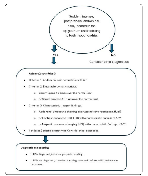
-Abdominal pain compatible with AP: acute onset of persistent, severe epigastric pain, often radiating to both hypochondria.
-Elevated serum lipase or amylase activity: at least three times greater than the upper limit of normal.
-Characteristic imaging findings on abdominal ultrasound and/or contrast-enhanced CT (CECT) and, in some cases, magnetic resonance imaging (MRI).
Gravity classification
Mild Acute Pancreatitis (MAP): absence of both (peri)pancreatic necrosis and organ failure (OF).
Moderately Severe Acute Pancreatitis (MSAP): presence of any type of sterile (peri)pancreatic necrosis and/or transient OF.
Severe Acute Pancreatitis (SAP): presence of any type of infected (peri)pancreatic necrosis or persistent OF.
Critical Acute Pancreatitis (CAP): presence of infected (peri)pancreatic necrosis and persistent OF (worse prognosis than SAP).
Transient OF: organ failure that resolves within 48 hours after appropriate supportive measures.
Persistent OF: organ failure that does not resolve within 48 hours despite appropriate supportive measures.
Peripancreatic collections are the most common local complications in AP. They may consist solely of inflammatory fluid or arise from necrosis and contain both solid and liquid components. (See Complication Handling)
Fluid collections related to acute pancreatitis in the early phase (< 4 weeks) are called acute peripancreatic fluid collections (APFC) and generally resolve spontaneously. After 4 weeks, these collections may become encapsulated and are then referred to as pseudocysts.
Collections related to necrotizing acute pancreatitis in the initial phase (< 4 weeks) are called acute necrotic collections (ANC), and if they persist for more than 4 weeks, they become encapsulated and are referred to as walled-off necrosis (WON).
Pancreatic necrosis
• (Peri)pancreatic necrosis is nonviable tissue located in the pancreas itself and/or in the peripancreatic area; it may be solid or semisolid (partially liquefied) and without a defined wall. It is detected by CECT, which reveals an area lacking contrast enhancement.
• Sterile (peri)pancreatic necrosis: absence of proven infection in the necrosis.
• Infected (peri)pancreatic necrosis: when at least one of the following signs is confirmed:
Table 2: Marshall criteria: the cutoff value is 2 or more, and OF may be transient (less than 48 hours) or persistent (more than 48 hours)
| MARSHAL SCORE. Complications | 0 | 1 | 2 | 3 | 4 |
| RESP. Pao2/FiO2 | 400 | 301-400 | 201 - 300 | 101 - 200 | 101 |
| RENAL | 1.4 | 1.4 - 1.8 | 1.9 - 3,6 | 3,7 - 4,8 | 4.9 |
| CARDIOVASCULAR. Systolic AP in Hg mm | 90 | 90 Responds to fluids | 90 Doesn’t respond to fluids | 90 Ph: 7.3 | 90 Ph: 7.2 |
Brahin FA, Suarez Anzorena Rosasco FJ. Estado actual del manejo de la pancreatitis aguda biliar. Rev Argent Cir. 2021;113 (Supl 2) Pag 127
Table 3: Balthazar tomographic criteria
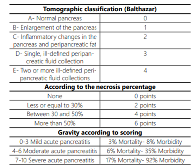
Fuente: Brahin FA, Suarez Anzorena Rosasco FJ. Estado actual del manejo de la pancreatitis aguda biliar. Rev Argent Cir. 2021;113 (Supl 2) Pag 129
1. Mild acute pancreatitis (MAP) is characterized by the absence of both (peri)pancreatic necrosis and organ failure.
2. Moderate acute pancreatitis (MoAP) is characterized by the presence of any type of sterile (peri)pancreatic necrosis or transient organ failure.
3. Severe acute pancreatitis (SAP) is characterized by the presence of any degree of infected (peri)pancreatic necrosis or persistent organ failure.
4. - Critical acute pancreatitis (CAP) is characterized by the presence of infected (peri)pancreatic necrosis and persistent organ failure.
Declarations and Recommendations
The diagnosis of AP requires two of the following three characteristics:
Abdominal pain is compatible with AP (acute onset of persistent, severe epigastric pain that often radiates to the back).
Serum lipase activity (or amylase activity) at least three times greater than the upper limit of normal.
Characteristic findings of AP on contrast-enhanced computed tomography (CT) and, less frequently, magnetic resonance imaging (MRI) or transabdominal ultrasound.
The most useful laboratory test for diagnostic confirmation, once AP is suspected, is serum lipase, whose elevation to twice the normal range has a sensitivity and specificity of about 95% for the diagnosis of AP.
Ultrasound is a valuable tool in the initial evaluation of AP; the most common cause is obstruction of the bile duct by gallstones, so ultrasound is the first imaging method to use, especially to identify the biliary cause and possibly detect some complications.
CT is suggested in:
Between the 3rd and 5th day of illness in AP.
In cases of diagnostic uncertainty and blunt abdominal trauma, during the same emergency consultation,
In the event of clinical deterioration.
When complications are suspected.
MRI is of usefulness due to its cost and availability. It can be useful for the follow-up of patients with gallstone-related AP and to evaluate the response to treatment, especially in children and patients allergic to contrast agents.
Regarding the severity classification in acute pancreatitis, it is recommended to use the Petrov classification or, failing that, the 2012 Atlanta classification.
For systemic complications, the Marshall classification provides an objective and accurate assessment for evaluating severity and making decisions.
SECTION 2: Initial Handling
Initial Handling
Initial handling is based on a combination of monitoring, supportive measures, pain medication, and management of complications, forming a multidisciplinary team.
Probiotics have been found to have a higher mortality rate in cases of severe AP and should therefore be omitted as part of treatment.
Antibiotic prophylaxis reduces infections in general but does not prevent pancreatic infection, pneumonia, urinary tract infection, complications, interventions, or mortality.
There is preliminary evidence indicating that Omega 3, short-chain fatty acids, and Infliximab modulate the inflammatory response in AP, but clinical benefits have not yet been demonstrated.
Fluid resuscitation
The acute phase of AP is characterized by inflammation and endothelial injury. There is third-space fluid loss, leading to tissue hypoperfusion.
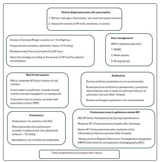
Adequate fluid replacement is crucial in the initial management of pancreatitis. An infusion rate of 1.5 ml/kg/h is recommended, with a bolus of 10 ml/kg if hypovolemia is present, and frequent reassessment every 6 hours during the first 24 hours. Severe AP is associated with higher fluid requirements and greater risk of complications, requiring closer monitoring. The strategy should be tailored to each patient and their comorbidities.
It has been reported that hydration with lactated Ringer’s solution is superior to normal saline, due to a lower risk of disease severity and a reduced ICU admission rate. Colloids have not demonstrated benefits in AP in ICU patients.
Nutritional support
In past decades, patients were kept fasting to allow the pancreas to “rest” and inflammation to subside. Recent clinical studies have shown that early oral nutrition promotes faster recovery, and shorter hospital stays in patients with mild or moderate AP. Very early oral feeding (<24 h) has not shown better outcomes than early feeding (>72 h). Enteral nutrition via feeding tube is recommended if oral intake is insufficient. There are no significant differences between nasogastric and nasojejunal feeding. When neither oral nor enteral feeding is possible due to persistent ileus, parenteral nutrition (PN) should be considered.
Pain management
Abdominal pain is the most common and distressing symptom in AP. Neither NSAIDs nor opioids have shown a significant impact on preventing disease progression in humans. The WHO step‑up approach is recommended, starting with non‑opioid analgesics such as NSAIDs, adding a weak opioid as the second step, and then strong opioids if needed. Renal impairment is a contraindication for NSAID use.
Antibiotics
After the period of intense inflammation, there is an anti‑inflammatory phase that can cause relative immunosuppression, increasing the risk of pancreatic and extra‑pancreatic infections. During the Systemic Inflammatory Response Syndrome (SIRS), fever may occur, and infection may be misdiagnosed, which is why in daily practice patients often receive antibiotics in the early phase (< 7 days).
Necrosis infection occurs mainly after 14 days. It is diagnosed by the presence of persistent clinical deterioration in the absence of other infections, the appearance of gas in pancreatic and peri‑pancreatic collections on imaging studies, or positive pancreatic tissue cultures.
Broad‑spectrum antibiotics are recommended for one to two weeks, or until a positive blood culture or pancreatic tissue culture is obtained, with therapy then directed according to the results. The use of broad‑spectrum antibiotics facilitates fungal infection; there are no studies indicating that antifungal prophylaxis is necessary.
Prokinetics
In patients with AP in the ICU, the administration of ondansetron was associated with better outcomes at 90 days. The use of ondansetron is recommended in ICU patients with nausea and vomiting.
Metoclopramide is a peripheral antagonist of dopamine (D2) receptors in the intestine. It also stimulates gastric emptying via muscarinic receptors. Neostigmine can be used to increase intestinal peristalsis and has been proposed for the treatment of colonic ileus associated with intra-abdominal hypertension that does not respond to basic treatments.
When intra-abdominal pressure exceeds 12 mmHg, the use of erythromycin and metoclopramide as prokinetics is recommended. In patients who do not respond, endoscopic decompression of the colon is recommended.
Neostigmine is an anticholinesterase drug that improves intestinal peristalsis; the latest studies published in March 2022 suggest that neostigmine may reduce intra-abdominal hypertension by promoting defecation. However, the clinical value for AP remains debatable, and current international guidelines do not place major importance on this drug.
Cholecystectomy
Cholecystectomy is a crucial step in gallstone-related AP. It should always be preceded by Magnetic Resonance Cholangiopancreatography (MRCP) or intraoperative cholangiography (IOC). Two systematic reviews and one multicenter clinical trial have shown that early cholecystectomy (during the same hospitalization) in patients with mild AP is safe and reduces the risk of complications such as recurrent pancreatitis and biliary colic compared with cholecystectomy performed after hospital discharge.
In moderate AP, surgery is delayed for 4 weeks after discharge to allow the inflammatory process to resolve before performing cholecystectomy.
In severe cases, it is recommended to wait until the inflammatory/infectious process has subsided before considering cholecystectomy, generally after 4 weeks.
Differential diagnosis
Differential diagnoses include perforated gastric or duodenal ulcer, mesenteric infarction, intestinal obstruction, aortic aneurysm, biliary colic, acute hepatitis, acute appendicitis, diverticulitis, inferior wall myocardial infarction, hematoma of the abdominal or splenic muscles, among others.
Declarations and Recommendations
Initial Handling:
Hydration with Ringer’s lactate. Moderate hydration: bolus of 10 ml/kg followed by infusion at 1.5 ml/kg/h. Frequent reassessment every 6 hours during the first 24 hours.
Do not use colloids.
If Ringer’s lactate is not available, use normal saline (0.9% sodium chloride).
Consider comorbidities to tailor hydration to each patient’s situation.
Antibiotics:
The use of antibiotics as prophylaxis in AP is not recommended.
In patients with infected necrosis, antibiotic therapy may avoid the need for intervention. Recommended antibiotics include carbapenems, quinolones, third-generation or higher cephalosporins, metronidazole (in combination with quinolones or cephalosporins). Vancomycin.
Fine-needle aspiration of infected necrosis is discouraged.
Antifungal therapy is indicated only when fungal infection is confirmed.
Procalcitonin is recommended as a guiding test for initiating, continuing, or discontinuing antimicrobial treatment.
Selective intestinal decontamination with neomycin or polymyxin is not recommended.
Pancreatin. Exocrine pancreatic insufficiency (EPI):
EPI is generally underdiagnosed in AP, but according to two meta-analyses its frequency is 27% to 30%.
EPI is secondary to inflammation, necrosis, infection, fibrosis, or ductal obstruction/disruption.
Diagnosis of EPI is based on clinical manifestations (abdominal distension, diarrhea, pain, presence of undigested food residues in the intestinal lumen), pancreatic secretion test, and fecal elastase, with values from 0 to 200 µg/g being suggestive of EPI, as well as nutritional parameters (biochemical and anthropometric).
Treatment of EPI: healthy oral diet or polymeric enteral nutrition, elimination of toxins (no cannabis, no alcohol, no smoking)
Enzyme replacement therapy: initially 6 capsules/day (one 25,000 IU capsule equals 300 mg of lipase)
Main meals: 2 capsules - Smaller meals: 1 capsule
In patients with severe pancreatitis or pancreatic necrosis > 50%, start enzyme replacement therapy. In mild pancreatitis with necrosis < 50%, confirm the diagnosis of EPI before starting treatment.
Prokinetics
The use of ondansetron has been associated with better 90‑day outcomes.
Prokinetic agents are frequently used in ICUs, although there is no strong evidence supporting their benefit.
Neostigmine was significantly more effective than conventional treatments in reducing intra‑abdominal hypertension in patients with AP. However, its clinical value for AP remains debatable according to current international guidelines.
The use of conventional prokinetics (ondansetron, metoclopramide, domperidone, levosulpiride) is left to the treating physician’s discretion, based on each patient’s symptoms.
- In cases of intra‑abdominal hypertension in ICU patients, consider the use of neostigmine.
Nutrition
It is important that the nutrition plan be individualized and supervised by a healthcare professional or nutritionist, as each patient’s needs may vary.
Enteral nutrition is preferable to parenteral nutrition whenever possible.
In mild AP, oral feeding should be started as soon as symptoms improve (abdominal pain, nausea, vomiting). Whenever possible, oral intake should not be interrupted.
In moderate and severe AP, the first option is oral feeding if tolerated; if after three days oral intake is not tolerated, a transpyloric tube should be placed and enteral nutrition initiated.
If nutritional intake does not meet minimum caloric requirements, or if enteral nutrition is not tolerated, parenteral nutrition is indicated.
Nutritional support is an essential component in the management of AP, and its approach should be individualized according to disease severity and patient tolerance.
In mild AP, the goal is to maintain continuous oral feeding, avoiding unnecessary interruptions. This highlights the importance of early refeeding to promote recovery and prevent complications.
In moderate and severe AP, oral feeding is recommended as the first option if tolerated, with enteral nutrition as the second option.
Parenteral nutrition is reserved for situations in which enteral nutrition is not tolerated or fails to meet the patient’s minimum caloric requirements.
Cholecystectomy
Early cholecystectomy in mild AP, together with prior IOC or MRCP, is the treatment of choice.
In moderate AP, cholecystectomy should be deferred until after 4 weeks from hospital discharge.
In severe AP, cholecystectomy with IOC or MRCP should be performed after 4 weeks from the resolution of complications.
Hospital protocols should be established to facilitate early cholecystectomy in patients with mild AP, including the availability of preoperative IOC and MRCP.
In patients with moderate acute pancreatitis, schedule cholecystectomy after 4 weeks from discharge, with preoperative IOC and MRCP.
SECTION 3: Acute Pancreatitis’ Complications
Acute Pancreatitis’ Complications
AP can present with a wide range of complications, which may be local or systemic, and may occur early or late. Peripancreatic necrosis stands out as a frequent complication and is a crucial indicator of disease severity. These can be:
Systemic complications
They originate from the systemic inflammatory response syndrome (SIRS) that accompanies AP. This uncontrolled inflammatory response can damage cells and release substances that cause vasodilation, increased vascular permeability, and edema, which can lead to serious complications such as acute respiratory distress syndrome (ARDS) and multiple organ failure (MOF).
Systemic complications can be very severe and even life‑threatening. There are two periods when the risk of death is highest:
Other possible systemic complications include:
Lungs: pleural effusion, atelectasis, ARDS.
Heart and blood vessels: low blood pressure, dehydration, pericardial effusion, thrombosis.
Renal: oliguria, hyperazotemia, thrombosis of the renal artery, vein, or both, acute tubular necrosis.
Metabolism: hyperglycemia, hypertriglyceridemia.
It is very important to detect and treat systemic complications in a timely manner.
It should be emphasized that systemic complications, in particular, are a complex issue that requires further study and an “interdisciplinary approach”. This consensus focuses mainly on complications requiring surgical treatment.
Local complications
Local complications of AP are managed differently depending on the composition of pancreatic and peripancreatic collections (pure fluid or necrotic solid content). Management of fluid collections requires precise timing, as the stage of evolution directly influences the therapeutic strategy. It is important to distinguish between:
Acute peripancreatic fluid collections (APFC): occur within the first four weeks and are typical of interstitial edematous AP. They usually appear in the first week, lack a defined wall, have a homogeneous internal structure, extend through peripancreatic spaces, and resolve spontaneously in most cases. Persistence beyond four weeks significantly increases the likelihood of evolving into a pseudocyst (PC).
Pseudocyst (PC): appears after four weeks. It is a fluid collection with a cyst-like appearance but with a fibrous wall instead of the epithelial lining of a true cyst, arising from an APFC more than four weeks after symptom onset.
Acute necrotic collection (ANC): develops within the first four weeks, involving both pancreatic parenchyma and peripancreatic tissues, with a variable mixture of fluid and solid material. The solid component helps distinguish ANC from APFC and PC.
When an area of necrosis is surrounded by a wall or capsule visible on imaging, it’s denominated Walled-off necrosis (WON). It develops from an ANC after four weeks from AP onset. The difference between WON and PC lies in the presence of variable amounts of solid material within the cavity.
Pancreatic necrosis: defined as non-viable tissue resulting from leakage of pancreatic fluid and immune cells, representing a form of tissue injury due to non-apoptotic cell death. Morphological changes are heterogeneous. Necrotic tissue appears as a lack of contrast enhancement in the affected pancreatic area, best visualized on CT scan after 72 hours from necrosis onset.
These complications are usually sterile at first but can become infected at any point during the course of the disease. They may be asymptomatic or symptomatic, causing compression of abdominal organs and, in some cases, systemic repercussions such as organ failure.
Other severe complications of surgical treatment
In these cases, the priority is to address the emergency, preserve the pancreatic compartment, and perform damage control for the findings encountered.
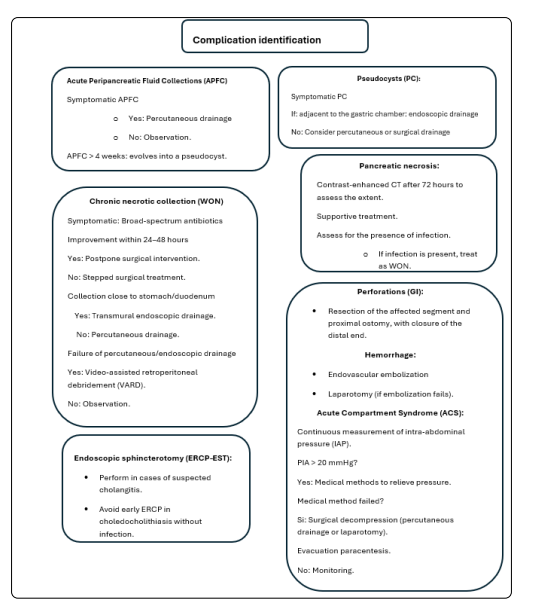
Perforations: rupture of the intestinal wall, either small bowel or colon, as a direct or indirect consequence of severe pancreatic inflammation. This is rare but extremely serious, with high mortality. In cases of colonic perforation, resection of the affected segment with proximal colostomy and closure of the distal stump is suggested.
Hemorrhage: bleeding may occur into the gastrointestinal tract or freely into the peritoneal cavity. Endovascular embolization is suggested, or laparotomy depending on hospital resources and patient support needs. Hemorrhagic complications such as splenic vein thrombosis or pseudoaneurysms may require surgical intervention if endovascular management fails.
Acute Compartment Syndrome (ACS): defined as sustained intra‑abdominal pressure (IAP) > 20 mmHg. Continuous IAP monitoring is essential for early detection. This is a severe complication with high mortality. When medical measures are insufficient to relieve IAP, surgical decompression via percutaneous drainage or laparotomy is indicated. Evacuative paracentesis can help reduce IAP.
Complications’ management
They will be managed according to the specific complication identified. All these complications, “as long as they remain asymptomatic”, do not require surgical intervention.
They become symptomatic when they exert a mass effect due to their size or become infected. It has been shown that the longer a collection evolves, the more likely it is to develop a wall and become better defined, which facilitates surgical treatment. On average, a waiting period of 4 weeks is recommended.
Pancreatic debridement should be avoided in the early acute phase (first 2 weeks), as it is associated with increased morbidity and mortality. Debridement should ideally be delayed for 4 weeks and performed earlier only when there is an organized collection and a strong indication.
APFC handling
Indication for drainage: compression of adjacent structures due to size, causing gastric outlet or biliary obstruction; disruption of the main pancreatic duct (Wirsung), infection, or bleeding. Percutaneous drainage is suggested.
Pseudocysts’ handling
Indication for drainage: persistent abdominal pain attributable to the pseudocyst, gastric, duodenal, or biliary obstruction, pancreatic ascites, progressive increase in size on imaging, infection, or hemorrhage.
The treatment of choice includes endoscopic, percutaneous, or surgical drainage. When selecting the technique, it is important to assess communication with the main pancreatic duct. Endoscopic transgastric approach (or transduodenal) drainage guided by endoscopic ultrasound is the most suitable when the pseudocyst is adjacent to the gastric chamber. This allows the creation of a cystogastrostomy without risk of spillage into the peritoneal cavity. The communication is established by placing stents, which are removed after a few weeks.
Percutaneous drainage is reserved for infected pseudocysts located far from the gastrointestinal lumen or when patient comorbidities contraindicate sedation or general anesthesia.
Surgical treatment is indicated when endoscopic treatment is contraindicated or has failed. Options include cystogastrostomy, cystoduodenostomy, or cystojejunostomy (preferably laparoscopic).
WON handling
Acute necrotic collections and “walled‑off” necrosis are usually sterile and resolve spontaneously with supportive treatment for AP. In the event of clinical deterioration, systemic toxicity, or suspected superinfection, some form of intervention is required. When there is no suspicion of infection, the indications for intervention include mechanical obstruction (gastric, intestinal, or biliary) and persistent symptoms for more than 8 weeks after the diagnosis of AP; when infection is documented or infection is suspected, surgical intervention should be indicated.
Indicators of infection of the necrosis are:
Clinical parameters (increased abdominal pain, fever >38.5 °C), laboratory parameters (white blood cells, CRP, or procalcitonin increasing), and imaging findings (increase in the collection and presence of air)
Persistence of sepsis despite correct intensive treatment
After having ruled out other possible sources of infection
New organ failure or persistence/worsening of the one already present.
Currently, fine needle aspiration (FNA) is not required to make the diagnosis of infection.
These patients should be evaluated daily by a multidisciplinary team. Upon making the diagnosis of infection, broad‑spectrum antibiotics (carbapenem, quinolones, metronidazole) are initiated for 24 to 48 hours; if the course is favorable, surgical intervention is postponed. If the course does not change or worsens, stepped surgical treatment is initiated.
The PANTER study has established stepped treatment as the standard in AP. Although most patients with AP do not require invasive intervention, there are special situations in which they may benefit.
Most local complications are resolved without invasive interventions. Acute peripancreatic collections generally resolve in 7-10 days. Only 6% of these collections develop into pancreatic pseudocysts, which tend to resolve spontaneously in up to 70% of cases. Drainage is indicated if, after 6 months of follow‑up, the patient develops symptoms such as abdominal pain, jaundice, early satiety, or fever, or if growth of the pseudocyst is demonstrated.
Endoscopic drainage
Indication: Collections close to the stomach or duodenum, accessible by endoscopy.
Endoscopic ultrasound guided transmural drainage is the preferred option. This procedure creates a tract between the collection and the gastric or duodenal lumen; it is performed under endoscopic visualization, placing a gastric transmural drainage of the double pig‑tail type or a metal stent. This metal stent, with a lumen wide enough, allows direct debridement of the necrotic material using endoscopic visualization and instruments.
Percutaneous drainage
Indication: collections distant from the gastrointestinal lumen, without access to endoscopic visualization.
Multifenestrated catheters are inserted under CT or ultrasound guidance toward the necrotic collection. CT offers better visualization of anatomical structures and detection of collections. Some centers use real‑time dynamic CT. Ultrasound allows the procedure to be performed at the bedside, without radiation, and at low cost. It can be combined with fluoroscopy in the operating room. Through these, lavages are performed, which must be systematically quantified. Lavages must meet aseptic criteria; 0.9% normal saline is recommended, or a preparation of 1000 cc of normal saline with 30 cc of povidone‑iodine solution and 30 cc of 10% hydrogen peroxide.
If the process is not controlled with these drains, progressive replacement of the catheters with others of larger size is recommended, as a tract for a future VARD.
Video-assisted retroperitoneal debridement (VARD)
Indication: when percutaneous or endoscopic drainage is technically not possible initially, or in case of failure of other previous treatments.
Necrosectomy, whether laparoscopic, open surgery, or a combination of both, is a major procedure that can trigger a significant inflammatory response in critically ill patients. Therefore, it is reserved for situations where minimally invasive strategies have failed or are not feasible. The matured and dilated tract created by prior percutaneous drainage is used.
Minimally invasive surgical approaches for the debridement of necrotizing AP are preferred over open surgical necrosectomy whenever possible, given the lower morbidity. Laparostomy is used only exceptionally.
Declarations and Recommendations
Multidisciplinary care: severe AP is a complex disease that requires interdisciplinary care. It is crucial to seek a medical center with a multidisciplinary team that includes gastroenterologists, surgeons, interventional radiologists, infectious disease specialists, intensive care specialists, nutritionists, and endocrinologists.
Local complications: it is recommended to use the definitions established in the 2012 Atlanta consensus.
It is recommended to base the necrosis infection diagnosis on the clinical and imaging signs that appear during the disease. FNA is not recommended for the diagnosis of infected necrosis.
Indications for intervention in the recommended complications:
Infection or superinfection of the necrosis
Symptoms that prevent a good course (respiratory difficulty or inability to receive oral feeding due to mass effect)
Step‑up multidisciplinary treatment, in evolutionary stages, has shown better results in the treatment of these patients
Controlled delay:
Delay surgical intervention until after the fourth week from the onset of the disease, as it is associated with better outcomes.
Constant monitoring of the patient is crucial to detect and treat complications in time.
Each patient must be evaluated individually, following the protocols and experience of each hospital.
Management of other surgical complications:
Conservative surgical approach: in complications such as hemorrhage or gastrointestinal perforations, surgery should focus exclusively on resolving the emergency, avoiding unnecessary interventions.
Special cases
Post-ERCP-EST acute pancreatitis
Post-ERCP AP is the most common major complication of ERCP, representing substantial morbidity and occasional death. In post-ERCP-EST pancreatitis with new abdominal pain, with elevation of pancreatic enzymes three times the upper limit of the normal range at 24 hours after the procedure, hospitalization for at least 48 hours is recommended.
Acute pancreatitis in pregnant women
AP in pregnant women is an uncommon but serious condition that can have significant consequences for both the mother and the fetus. During pregnancy, hormonal and physical changes can increase the risk of this condition.
The most common causes of AP during pregnancy are the same as in non-pregnant women. It is crucial to seek immediate medical attention if symptoms of AP occur during pregnancy. Symptoms may be nonspecific.
Early diagnosis and treatment can significantly improve outcomes for both the mother and the baby. Severe AP can lead to serious complications such as preterm delivery, preeclampsia, acute respiratory distress syndrome (ARDS), infection, and, in severe cases, it can be fatal.
Acute pancreatitis in children
AP is less common in children than in adults, but it can be severe and require immediate medical attention. The causes in children can vary and are often not easily identified. It is essential to seek immediate medical attention if a child presents symptoms of pancreatitis. Early diagnosis and treatment can prevent serious complications.
Preventive measures
(Risk factors’ control)
Maintain a healthy diet: following a low‑fat diet rich in fruits, vegetables, and whole grains helps prevent the formation of gallstones.
Limit alcohol consumption: reducing or eliminating its intake can significantly decrease the risk.
Control weight: maintaining a healthy weight through a balanced diet and regular exercise is essential to prevent cardiovascular events as well as the formation of gallstones.
Do not smoke: smoking increases the risk of pancreatitis as well as many other neoplastic diseases.
Treat underlying conditions: some medical conditions, such as gallstones and hypertriglyceridemia, can increase the risk of pancreatitis. Treating these conditions can help prevent AP.
Increase water intake: staying hydrated is important for overall health and can also help prevent AP.
Engage in regular physical exercise: moderate or high‑intensity regular physical activity can help maintain a healthy weight and reduce the risk of gallstones.
If you have a family history of AP or experience symptoms such as severe abdominal pain, nausea, or vomiting, it is important to consult a doctor. Early diagnosis and treatment can help prevent complications.
Conclusions
The consensus establishes a reference framework based on scientific evidence, adapted to the particularities of the Paraguayan national context. This allows for homogeneous clinical practice.
The adoption of standardized classifications and protocols facilitates communication among health professionals and improves decision-making.
Acute biliary pancreatitis is the most frequent cause of AP in Paraguay.
The implementation of the consensus recommendations would result in comprehensive and adequate care for patients with AP, which translates into improved outcomes.
A multidisciplinary approach is promoted, involving specialists in gastroenterology, surgery, intensive care, infectious diseases, interventional procedures, and nutrition, to ensure complete and coordinated care.
Early identification of risk factors and the implementation of appropriate initial treatment will help reduce the severity of AP and prevent complications.
The consensus promotes clinical research in key areas, generating local evidence that allows the adaptation of clinical practice guidelines to the Paraguayan reality and thus improves patient care.
The consensus recognizes the importance of continuous training for health professionals, especially general surgeons, gastroenterologists, and primary care physicians. The organization of courses, workshops, and updating activities are encouraged to disseminate the consensus recommendations and improve the competence of professionals in the management of AP.
In summary, the consensus on the management of acute pancreatitis in Paraguay represents a significant step forward in improving the care of this disease. The implementation of the consensus recommendations and the promotion of clinical research are essential to ensure that all patients receive quality care and to reduce the complications and mortality associated with AP.
Limitations
Accessibility and applicability:
Limited resources:
Some may require technology or treatment recommendations that are not widely available in all hospitals in Paraguay, especially in rural or less-resourced areas.
The availability of specialized intensive care units or percutaneous drainage procedures may be limited.
Variability in implementation:
Despite efforts to unify criteria, the implementation of the consensus may vary among different hospitals and health professionals.
Factors such as lack of training, resistance to change, or work overload may hinder the adoption of the recommendations.
Evidence and local context:
Research gaps:
The available scientific evidence may not be fully applicable to the Paraguayan context, due to differences in epidemiology, risk factors, or population characteristics.
The lack of local studies may limit the ability to adapt the recommendations to the Paraguayan reality.
Changes in evidence:
Medicine is constantly evolving, and new research may modify or refute the recommendations of the present consensus.
It is necessary to periodically update the consensus to incorporate the latest scientific advances.
Socioeconomic and cultural factors:
Barriers for access:
Factors such as poverty, lack of medical coverage, or cultural barriers can hinder patients’ access to timely medical care.
Delays in diagnosis and treatment can increase the severity of AP and worsen outcomes.
Adherence to treatment:
Cultural differences or health beliefs can influence patients’ adherence to treatment and preventive recommendations.
Examples:
The recommendation to perform percutaneous drainage of infected pancreatic necrosis may be difficult to implement in hospitals that do not have trained interventional radiologists.
Recommendations on early enteral nutrition may be difficult to follow in patients with limited resources or with difficulties accessing specialized foods.
Lack of awareness of the risks of alcohol consumption in rural areas, where the consumption of alcoholic beverages is more common.
It is important to recognize these limitations and work to overcome them through the training of professionals, improvement of health infrastructure, promotion of local research, and promotion of the present consensus.
Recommendations for the implementations of the consensus
Dissemination and education:
Continuous training:
Conduct workshops, courses, and seminars aimed at general practitioners, surgeons, intensivists, nurses, and other health professionals involved in the management of AP.
Use online platforms and educational materials to ensure access to information for all professionals, even in remote areas, for example, the MSP and BS website.
Support materials:
Develop clinical practice guidelines, management protocols, and decision-making algorithms based on the consensus, adapted to the Paraguayan context.
Create support tools, such as severity classification tables, risk scales, and follow-up sheets, to facilitate the application of the consensus in daily clinical practice and distribute them in MSP and BS facilities.
Public awareness:
Carry out information campaigns aimed at the general population to increase awareness of risk factors, symptoms, and the importance of early diagnosis of AP.
Implementation and Follow-up:
Adaptation to the local reality:
Consider the resource limitations and the characteristics of the Paraguayan health system when implementing the consensus recommendations.
Establish priorities and strategies for gradual implementation, focusing on interventions with the greatest clinical impact and cost-effectiveness.
Audit and feedback:
Establish quality indicators to assess the implementation of the consensus and its impact on clinical outcomes.
Conduct periodic audits to identify areas for improvement and provide feedback to healthcare professionals.
Collaboration networks:
Establish networks among hospitals and referral centers to facilitate the transfer and care of patients with complications.
Promote communication and coordination among the different levels of care to ensure continuity of care.
Promote clinical research evidence on the in Paraguay to generate epidemiology, risk factors, and effectiveness of therapeutic interventions in the local population.
Establish patient registries and databases to facilitate research and long-term follow-up.
Periodically review and update the consensus as new scientific evidence becomes available and changes in clinical practice occur.
Maintain fluid communication with the international scientific community to incorporate the latest advances in the management of AP.
Coordination between the Ministry of Public Health and Social Welfare, the Paraguayan Society of Surgery (SOPACI), and the Paraguayan Hepato-Pancreato-Biliary Chapter is necessary for the dissemination and implementation of the recommendations.
These algorithms are a guide and should be used in conjunction with clinical judgment and must be adapted to each individual patient.
Bibliografía
1. Ausania F, Senra del Río P. Estrategias terapéuticas de la pancreatitis aguda y sus complicaciones. Guías clínicas de la Asociación Española de Cirugía. 2da edición. Cirugía Biliopancreatica. Cap 13. ARAN. 2018. Pág 201- 221 [ Links ]
2. Banks PA, Bollen TL, Dervenis C, Gooszen HG, Johnson CD, Sarr MG, et al. Clasificación de la pancreatitis aguda - 2012: revisión de la clasificación de Atlanta y definiciones por consenso internacional. Intestino. 2012;62(1):102-11. doi:10.1136/gutjnl-2012-302779. [ Links ]
3. Bakker OJ, van Santvoort HC, van Brunschot S, Geskus RB, Besselink MG, Bollen TL, et al. Minimally invasive 'step-up approach' versus maximal necrosectomy in patients with acute necrotising pancreatitis (PANTER trial): design and rationale of a randomised controlled multicenter trial [ISRCTN13975868]. BMC Surg. 2006 Apr 11;6:6. doi: 10.1186/1471-2482-6-6 [ Links ]
4. Baron TH, DiMaio CJ, Wang AY, Morgan KA. American Gastroenterological Association Clinical Practice Update: Management of Pancreatic Necrosis. Gastroenterology. 2020 Jan;158(1):67-75.e1. doi: 10.1053/j.gastro.2019.07.064 [ Links ]
5. Beger HG, Rau B, Mayer J, Pralle U. Natural course of acute pancreatitis. World J Surg. 1997;21(2):130-5. [ Links ]
6. Beij A, Verdonk R, van Santvoort H, de Madaria E, Voermans R. Pancreatitis aguda: una actualización del tratamiento basado en evidencia y tendencias recientes en estrategias de tratamiento. United European Gastroenterol J. 2025;00:1-10. https://doi.org/10.1002/ueg2.12743 [ Links ]
7. Brahin FA, Suarez Anzorena Rosasco FJ. Estado actual del manejo de la pancreatitis aguda biliar. Rev Argent Cir. 2021;113(Supl 2):1-219. [ Links ]
8. Boadas J, Balsells J, Busquets J, Codina-B A, et al. Assessment and treatment of acute pancreatitis. Position document of the Catalan Society of Gastroenterology, Catalan Society of Surgery and Catalan Society of the Pancreas. Gastroenterol Hepatol. 2015;38(2):82-96. doi:10.1016/j.gastrohep.2014.09.006. [ Links ]
9. Buter A, Imrie CW, Carter CR, Evans S, McKay CJ. Dynamic nature of early organ dysfunction determines outcome in acute pancreatitis. Br J Surg. 2002;89(3):298-302. [ Links ]
10. Conwell DL, Bank P, Greenberger NJ. Pancreatitis Aguda y Crónica. En: Kasper DL, Hauser S, Jameson L, Fauci AS, Longo D, Loscalzo J, editors. Harrison Principios de Medicina Interna. 19a ed. México DF; 2016. pp. 2090-8. [ Links ]
11. Dellinger EP, Forsmark CE, Layer P, Lévy P, Maraví-Poma E, Petrov MS, et al. Determinant-based classification of acute pancreatitis severity: An international multidisciplinary consultation. Ann Surg. 2012;256(6):875-880. doi:10.1097/SLA.0b013e318256f778. [ Links ]
12. Greenberg JA, Hsu J, Bawazeer M, Marshall J, Friedrich JO, Nathens A, et al. Clinical practice guideline: Management of acute pancreatitis. Can J Surg. 2016;59(2):128-140. doi:10.1503/cjs.015015 [ Links ]
13. Guilabert L, Cárdenas K, de Madaria E. Tratamiento inicial de la pancreatitis aguda. Gastroenterol Clin North Am. 2025; 54:21 36. https://doi.org/10.1016/j.gtc.2024.07.001 [ Links ]
14. Muñoz D, Medina R, Botache WF, Arrieta RE. Acute pancreatitis: Key points. Argumentative review of the literature. Rev Colomb Cir. 2023;38(2):339-351. doi:10.30944/20117582.2206. [ Links ]
15. Ocampo C, Kohan G, Leiro F, Basso S, Gutiérrez S, et al. Hospital Interzonal Eva Perón. Acta Gastroenterol Latinoam. 2015;45(4). Disponible en: www.actagastro.org [ Links ]
16. Puentes AB, Prieto Hernández JA. Pancreatitis Aguda. Artículo de Revisión. Intramed J. 2014; 3(2):1-14. [ Links ]
17. Purschke B, Bolm L, Meyer MN, Sato H. Interventional strategies in infected necrotizing pancreatitis: Indications, timing, and outcomes. World J Gastroenterol. 2022;28(27):3383-3397. doi: 10.3748/wjg.v28.i27.3383. [ Links ]
18. Villalba Aucejo S, Alfonzo Ramos A, Acuña Macchi J, Penner Sawatzky D. Albúmina y PCR como predictores de mortalidad en pacientes con pancreatitis aguda. ? Cátedra de Fisiopatología, Facultad de Ciencias Médicas. ? Universidad Nacional de Asunción, Paraguay. ? 2018. [ Links ]
19. Wu G, Ma Y, Wei W, Zeng J, Han Y, Song Y, et al. Ondansetron: recommended antiemetics for patients with acute pancreatitis? a population-based study. Front Pharmacol. 2023 May 10;14:1155391. doi: 10.3389/fphar.2023.1155391. PMID: 37234720; PMCID: PMC10205993. [ Links ]
20. Zarnescu NO, Dumitrascu I, Zarnescu EC, Costea R. Abdominal Compartment Syndrome in Acute Pancreatitis: A Narrative Review. Diagnostics (Basel). 2022 Dec 20;13(1):1. doi: 10.3390/diagnostics13010001. PMID: 36611293; PMCID: PMC9818265. [ Links ]
Received: March 28, 2025; Accepted: April 11, 2025
 Este es un artículo publicado en acceso abierto bajo una licencia Creative Commons
