




Serviços Personalizados
Journal

Artigo
 texto em
 Inglês (pdf)
 Artigo em XML
 Referências do artigo

 Tradução automática
 Enviar este artigo por email
Indicadores
-
 Citado por SciELO
Links relacionados
-
 Similares em
SciELO
Compartilhar

 Permalink

Cirugía paraguaya
versão On-line ISSN 2307-0420
Cir. parag. vol.49 no.1 Asunción abr. 2025
https://doi.org/10.18004/sopaci.2025.abril.20
Case Report
Acute pancreatitis and retroperitoneal perforation after endoscopic retrograde cholangiopancreatography: case report
1
 http://orcid.org/0000-0002-4842-4975
1
http://orcid.org/0009-0000-0898-0654
1
http://orcid.org/0009-0008-5730-0502
1Hospital de Clínicas, Primera Cátedra de Clínica Quirúrgica. San Lorenzo. Paraguay
A 54-year-old patient underwent ERCP for choledocholithiasis and developed severe acute biliary pancreatitis and a type II retroperitoneal perforation. Diagnosis was confirmed by imaging and managed with percutaneous drainage and conservative treatment, with good clinical evolution. This case highlights the importance of early recognition of serious ERCP complications and the success of non-surgical management.
Keywords: ERCP; Acute biliary pancreatitis; Retroperitoneal perforation; Post-endoscopic complications; Percutaneous drainage
Paciente de 54 años sometida a Colangiopancreatografía Retrógrada Endoscópica (CPRE) por coledocolitiasis, que desarrolló pancreatitis aguda biliar severa y perforación tipo II retroperitoneal. El diagnóstico se confirmó por imagen y se manejó con drenaje percutáneo y tratamiento conservador, con buena evolución clínica. El caso destaca la importancia del reconocimiento precoz de complicaciones graves post-CPRE y el éxito del manejo no quirúrgico.
Palabras claves: CPRE; Pancreatitis aguda biliar; Perforación retroperitoneal; Complicaciones postendoscópicas; Drenaje percutáneo
INTRODUCTION
Endoscopic retrograde cholangiopancreatography (ERCP) is an advanced endoscopic procedure in which an endoscope is guided to the duodenum, allowing instruments to pass through the ampulla of Vater into the bile ducts and the pancreatic duct. The ducts are opacified by injecting contrast medium, and radiographic methods are used to obtain images and facilitate endoscopic therapy. ERCP-guided procedures have been used to manage a variety of pancreatobiliary disorders.
It is a comprehensive procedure that requires special training and experience and is associated with a higher risk of adverse events than other endoscopic procedures. For this reason, it is important to use strategies to prevent adverse effects, to recognize them early, and to treat them if they occur1.
Reports on adverse events related to ERCP indicate a rate of 5 to 12% and a mortality rate of 0.1 to 1.4%. Among the risk factors, some may be related to sedation or to the endoscopy itself, or to an intervention performed during it. Cited factors include those related to the procedure (such as difficult cannulation), patient-related factors (anatomical alteration, sphincter of Oddi dysfunction, periampullary diverticulum, cirrhosis, advanced age), and factors related to the hospital and endoscopist (a low ERCP volume, procedure time, and selected cases such as acute cholangitis)1.
The most common adverse effects are pancreatitis, bleeding, perforation, and infection. Anesthesia-related effects have also been reported.
Pancreatitis is a very serious complication of ERCP, which may result from mechanical injury to the pancreatic duct, hydrostatic injury from contrast injection, or manipulation with the guide2. The incidence of post-ERCP pancreatitis is 3.5 to 9.7%. Pancreatitis can be severe in 0.3 to 0.8% of cases3.
Bleeding during ERCP is typically related to instrumentation and sphincterotomy. The incidence is around 1 to 3% (4). Infection after the procedure may be related to incomplete drainage of the bile duct, obstruction of the cystic duct, an infected pancreatic fluid collection, or, rarely, contamination of the endoscopic equipment. The prevalence rate is estimated at 3% or more5.
Post-ERCP perforation can occur in the esophagus, stomach, or small intestine. They can be classified according to Stapfer by mechanism as follows: Type I, due to perforation of the duodenal wall; Type II, retroperitoneal duodenal perforation secondary to periampullary injury; Type III, perforation of the main pancreatic duct; and Type IV, presence of gas in the retroperitoneum. Type II is the most common form6.
CLINICAL CASE
Female patient, 54 years old, cholecystectomized 6 years ago, was admitted after referral from an outpatient clinic with a one-month history of episodes of colicky pain in the right hypochondrium, non-radiating, related to the intake of fatty foods, partially relieved with the use of Lysine Clonixinate with Propinox; 24 hours prior to admission, the patient reported dark urine. She denied fever, jaundice, acholia/hypocholia, as well as nausea and vomiting.
History of the current illness: previously admitted to the service for a similar condition; on that occasion, complementary studies were performed and she was discharged with plans to undergo Magnetic Resonance Cholangiography (MRC) due to the absence of significant findings on abdominal ultrasound and laboratory tests within normal ranges. The MRC reported choledocholithiasis (dilation of the intrahepatic and extrahepatic bile ducts, more pronounced at the level of the common hepatic duct and the common bile duct, where it measured 21 mm); at this level, a hypointense image was observed causing an endoluminal filling defect of 9.6 mm.
ERCP was performed, revealing a juxtapapillary diverticulum. Cannulation of the common bile duct was achieved, contrast was injected, and the common bile duct was visualized with an approximate diameter of 20 mm and a filling defect in the distal third. A sphincterotomy with guided papillotomy and dilation with a hydrostatic balloon was then performed without immediate complications, followed by biliary exploration with a Dormia basket on several occasions. As a result of this procedure, one stone was successfully extracted. Table 1 shows the most relevant laboratory data from the post-procedure follow-up.
Table 1. Post-ERCP laboratory data.
| HB/HTO | 14.7/43.8% |
| GB/N | 10460/74% |
| Amylasemia | 102 |
| BT/BD/BI | 0,91/0,46/0,45 |
| AST/ALT/FA | 94/101/94 |
On the first day after the procedure, the patient reported severe abdominal pain that did not respond to medication, leading to the diagnosis of mild biliary acute pancreatitis without jaundice according to the Atlanta Classification. On the same day, abdominal ultrasound and non-contrast computed tomography (CT) of the abdomen and pelvis were performed due to suspected perforation, revealing a small amount of free fluid in the abdomen without direct signs of perforation. Table 2 and Table 3 show the most notable changes 48 and 72 hours after the procedure.
Table 2. Laboratory data 48 h Post-ERCP
| HB/HTO | 14.2/44% |
| GB/N | 3690/77% |
| Amylasemia | 172 |
| BT/BD/BI | 0,96/0,59/0,38 |
| AST/ALT/FA | 46/79/68 |
| Lipasemia | 274 |
| Platelets | 170000 |
Table 3. Laboratory control on the third post-procedure day
| HB/HTO | 14.2/44% |
| GB/N | 2720/79% |
| BT/BD/BI | 0,96/0,59/0,38 |
| AST/ALT/FA | 46/79/68 |
| Platelets | 63000 |
| pH | 7.32 |
| Pco2 | 33 |
| P02 | 65 |
| EDB | -9 |
| Bicarbonate | 17 |
| Saturation | 90% |
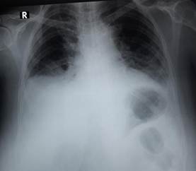
According to the Marshall criteria, a diagnosis of severe biliary acute pancreatitis with multiple organ failure was made, for which the patient was transferred to the ICU. During hospitalization, diagnoses of NIH and Chikungunya were also made. A standing chest X-ray was requested to rule out pneumoperitoneum (see Figure 1).
Despite improvement, suspicion of post-ERCP perforation persisted, so a contrast-enhanced (IV and oral) CT scan of the abdomen and pelvis was performed, revealing a collection in the right paracolic gutter (see Figure 2), extending to the right iliac fossa (RIF), without contrast leakage into the cavity, with a collection in the right paracolic gutter; the calculated volume was 192 ml. Percutaneous drainage was performed as treatment for this collection.
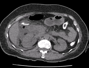
Figure 2. CT scan of the abdomen and pelvis with intravenous (IV) and oral contrast. Right pararenal retroperitoneal collection.
A follow-up study was performed, confirming the right pararenal retroperitoneal collection and a hypodense area in the posterior wall of the second portion of the duodenum, interpreted as a wall defect visible in Figure 3. A duodenoscopy was then carried out up to the second portion, revealing CO₂ filling of the collection bag corresponding to the percutaneous drainage, which indicated the presence of a perforation.
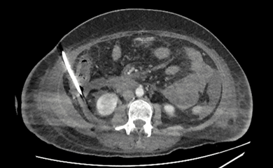
Figure 3. CT scan of the abdomen and pelvis with intravenous (IV) and oral contrast. Image of collection after percutaneous drainage.
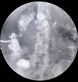
The patient received nutrition via nasojejunal tube (NJT), later progressing to an oral diet, as well as broad-spectrum antibiotic therapy: Meropenem® 1 g every 8 hours, Sodium colistimethate (Colistin®) 100 mg every 8 hours, and Tigecycline® 50 mg every 12 hours for 14 days. After 40 days of hospitalization, the patient was discharged in good condition, with plans to remove the drain in the following weeks. A fistulography was performed 39 days after drain placement, confirming an enteroatmospheric fistula (Figure 4), for which closure was decided. The drain was removed without complications one week later.
DISCUSSION
Post-ERCP complications can be mild or severe and require close clinical monitoring. In this case, the patient reported two of the most relevant complications described in the literature: acute biliary pancreatitis and type II retroperitoneal perforation.
The incidence of post‑ERCP pancreatitis reported in international studies ranges from 3.5% to 9.72, and its course can vary from mild forms to severe presentations with multiorgan failure, as observed in this patient. In the national context, a study conducted at the Hospital Nacional de Itauguá reported an incidence of 3.4%, which is consistent with international data, but most cases did not progress to severe forms7.
Type II perforation, secondary to a periampullary injury, is the most common form of post‑ERCP perforation. It is generally associated with sphincterotomy and other instrumental endoscopic maneuvers6. In this case, the diagnosis was initially suspected based on imaging findings and confirmed by duodenoscopy, which allowed the initiation of conservative management with percutaneous drainage, antibiotic therapy, and nutritional support.
The non‑surgical treatment was successful, in line with current recommendations for type II perforations, which tend to respond well to conservative management when detected early (8) . Multidisciplinary follow‑up, intensive care unit support, and the availability of high‑resolution imaging were key factors in achieving a favorable outcome.
This case highlights the importance of early recognition of the clinical and biochemical signs of post‑ERCP complications, as well as the need to perform imaging studies when there is clinical suspicion. Prompt and appropriate intervention can prevent surgical procedures and improve prognosis.
In conclusion, post‑ERCP monitoring must be rigorous, especially in patients with anatomical risk factors such as a juxtapapillary diverticulum. The treating team’s experience, the availability of diagnostic and therapeutic resources, and close follow‑up are fundamental pillars for the successful resolution of these complications.
REFERENCES
1. Tringali A, Costamagna G. UpToDate. 2023 [citado 10 de febrero de 2024]. Adverse events related to endoscopic retrograde cholangiopancreatography (ERCP) in adults - UpToDate. [ Links ]
2. Tringali A, Costamagna G. UpToDate. 2023 [citado 21 de junio de 2024]. Overview of endoscopic retrograde cholangiopancreatography (ERCP) in adults -UpToDate . [ Links ]
3. Tringali A, Costamagna G. UpToDate. 2023 [citado 21 de junio de 2024]. Post-endoscopic retrograde cholangiopancreatography (ERCP) pancreatitis -UpToDate . [ Links ]
4. Tringali A, Costamagna G. UpToDate. 2023 [citado 21 de junio de 2024]. Post-endoscopic retrograde cholangiopancreatography (ERCP) bleeding -UpToDate . [ Links ]
5. Tringali A, Costamagna G. UpToDate . 2022 [citado 21 de junio de 2024]. Infectious adverse events related to endoscopic retrograde cholangiopancreatography (ERCP) -UpToDate . [ Links ]
6. Tringali A, Costamagna G. Post-ERCP perforation - UpToDate [Internet]. 2022 [citado 10 de febrero de 2024]. [ Links ]
7. Aranda M, Prieto G, Petty-Bottin C, Adorno C. Scielo. 2022 [citado 21 de junio de 2024]. Incidencia de complicaciones precoces de la colangiopancreatografía retrógrada endoscópica en el Hospital Nacional de Itauguá. Periodo 2013-2017. [ Links ]
Conflict of interest: The authors declare that they have no conflicts of interest related to the preparation of this manuscript.
Received: January 05, 2025; Revised: January 20, 2025; Accepted: February 09, 2025
 Este es un artículo publicado en acceso abierto bajo una licencia Creative Commons
