




Serviços Personalizados
Journal

Artigo
 texto em
 Inglês (pdf)
 Artigo em XML
 Referências do artigo

 Tradução automática
 Enviar este artigo por email
Indicadores
-
 Citado por SciELO
Links relacionados
-
 Similares em
SciELO
Compartilhar

 Permalink

Cirugía paraguaya
versão On-line ISSN 2307-0420
Cir. parag. vol.48 no.2 Asunción ago. 2024
https://doi.org/10.18004/sopaci.2024.agosto.29
Case report
Volvulus of the right colon in adults with infantile cerebral paralysis
1
 http://orcid.org/0000-0003-4286-3931
1
http://orcid.org/0009-0001-8794-5482
1Centro Médico Bautista. Asunción, Paraguay
Two cases of volvulus of the cecum and right colon are reported in adults with Cerebral Paralisys. A 34-year-old man and a 48-year-old woman - were hospitalized for intestinal occlusion of 1 and 2 days of evolution. In both cases, the interrogation and physical examination contributed little or nothing, except for abdominal pain. The diagnosis of colon volvulus required imaging studies. At the time of surgery both had necrosis and the right colectomy was performed with ileostomy and mucosal fistula. Postoperative problems and mainly a slow gastrointestinal recovery postponed discharge.
Key words: volvulus of the right colon; Infantile cerebral paralysis
Se reportan dos casos de vólvulo del ciego y colon ascendente en adultos portadores de parálisis cerebral infantil. Un varón y una mujer de 34 y 48 años fueron hospitalizados por oclusión intestinal de 1 y 2 días de evolución; en ambos casos el interrogatorio y el examen físico aportaron poco o nada, excepto el dolor abdominal; el diagnóstico del vólvulo del colon requirió de estudios por imágenes. Al momento de la intervención ambos tenían necrosis y se les practicó la colectomía derecha con Ileostomía y fístula mucosa; diversos problemas postoperatorios y fundamentalmente una recuperación gastrointestinal lenta postergaron el alta.
Palabras clave: vólvulo del colon; Parálisis cerebral infantil
INTRODUCTION
Volvulus of the right colon constitutes an unusual cause of acute colon occlusion, much less frequent than sigmoid colon; the most mentioned causes are constipation, dolichocolon, and chronic consumption of laxatives1.
The distinctions between the volvulus of the cecum and volvulus of the colon are imprecise; the resulting differences are: precise anatomical limit and rotation axis; in any case the terminal ileum is (by rule) affected in lesser or greater measure resulting in some authors grouping it with the ileocecocolic volvulus denomination2. In the case of the “characteristic” volvulus of the right colon, a rotation above the ascending mesocolon, cecum and terminal ileum can be proven; the incomplete binding of the ascending colon is the constant anatomical condition, being ischemia and later necrosis a constant due to, among others, regularly a late diagnosis. The diagnosis is mainly clinical, however, the characteristic radiological findings in simple X-rays, ultrasound and CT scan help differentiate from other intestinal occlusion causes3.
The incapacitating neurological affections such as volvulus’ causes aren’t frequently reported. In these patients, however, early diagnosis turns particularly difficult due to scarce interrogatory input from the patient, just as physical exam and imagery studies’ limitations: anatomy alterations, mostly limb rigidness, dorsal-lumbar column deformity and spastic musculature4.
Infantile cerebral palsy (ICP) is a chronic disorder, non-progressive, of movement, posture and muscular tone, secondary to injuries in the central nervous system during early stages of life. Even though the fundamental alteration which these patients present is neurological, their implications encompass practically every apparatus and system. Nearly every ICP patient presents gastrointestinal symptoms and/or alterations to their nutritional state at some point in their lives5.
The objective of the present study is to illustrate the proven struggles of 2 ICP patients in early diagnosis and postoperatory handle, in the Bautista Medical Center (BMC).
CASE PRESENTATION
First case
34-year-old male, known ICP patient, sent for general state decay, feverish feeling and abdominal pain of 24hs of evolution. Admitted into said center due to suspicion of acute abdomen is discharged with alleviated pain. Reports again proving a urinary tract infection, given ciprofloxacin and ceftazidime; is transferred to BMC due to lack of betterment.
Stable, afebrile, nauseous flatulence and negative catharsis upon admission in the last 5 days ago (data given by family members). Soft abdomen, no defenses, normal bowel sounds. On the 4th day of admission, abdominal pain persists which encompasses all quadrants; an abdomen X-ray yields colon dilation, and abdomen and pelvis CT scan reveals large ascending colon dilation (12 cm), bowel sounds, with obstruction signs, with images suggesting mesenteric vascular vortex (Figure 1) compatible with volvulus of the cecum. Furthermore, CT scan yields spastic diplegia with generalized muscular atrophy, column with major thoracolumbar levoconvex scoliosis. Preoperatory studies yield 56%, proteinemia: 4,7 g/dL, albumin 2,7%, ASA III.
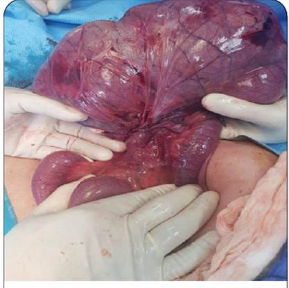
Volvulus of the cecum and ascending colon with necrosis are discovered during operation (Figure 2); a right hemicolectomy is performed, with ileostomy and transverse-level mucous fistula. Patient is admitted into intensive care unit; the ileostomy’s debit is null in the following 48-hours, while gastric residue is bilic and gradually increases, adding nausea. Patient is submitted to an abdomen X-ray which shows bowel sounds. Parenteral nutrition is started on the 3rd day.
Due to unproper evolution, surgical reinvention is decided upon and a large quantity of aching peritoneal liquid and secondary occlusion to an internal hernia (herniated thin loop in a breach of the transverse mesocolon), with dilation next to the flaps. A reduction in the strangulated closed-loop and closure of the aforementioned breach are performed.
Within the intensive care unit 48-hours after reintervention, no ileostomy’s debit continues, therefore touching through ostomy is performed, proving a 90% stenosis. During the third intervention, clear peritoneal liquid within the cavity and moderated dilation of the loops from the ostomy up to Treitz’s angle are discovered. Due to a lacking parietal orifice, it’s opted to dissect deep muscular plane (transverse) in a 3 to 4 cm extension.
Patient presents proper postoperatory initial evolution, although with ostomy level’s null debit and average debit through nasogastric tube of 2000 cc in 24-hours. On the fifth day of the third intervention it’s evaluated by a coloproctologist, who after performing touch at the ileostomy’s level ascertains an adequate orifice and advices insisting with conservative handle. Two days later, the patient remains without ileostomy debit and is drained by nasogastric tube 2000 cc in 24-hours; patient is evaluated by a different surgeon at the family’s behest, who after performing touch concludes that the ostomy’s parietal orifice is narrow, requests a CT scan and recommends reintervention, which is scheduled for the next day.
During the early morning, the ileostomy sharply drains 1000 cc of intestinal liquid, followed by a progressive decrease in the drainage through the nasogastric tube. In the following days, proper tolerance of the oral way is proven, and the patient is discharged from BMC.
Pathological anatomy reports: small intestine’s segment and colon with ischemic changes secondary to the volvulus of the cecum. No neoplastic process observed.
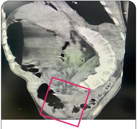
Figure 1. Simple CT scan of the abdomen and pelvis. Sagittal section with “vortex” imagery in the right iliac fossa.
Second case
48-year-old female, ICP patient, bedridden from birth, with records of seizure during regular treatment with carbamazepine and clonazepam, reports to emergency services due to a 48-hour evolutive case of lack of appetite, stool retention, abdominal pain and distention and vomiting. The exam reports pronounced abdominal distention and increased bowel sounds; abdomen X-rays show pronounced colon dilation.
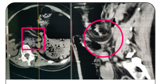
Patient is awake, uncooperative, with expressive aphasia, spastic tetraplegia, hypertonia. Spinal column scoliosis. Abdomen X-ray and CT scan show intestinal loop’s distension with presence of residue levels across the entire colon in great quantities; a change of direction of the mesenteric vessels can be seen in the transversal and sagittal sections (Figure 3). Upon touching, hardened stools are discovered and enemas are indicated. Patient doesn’t show clinical betterment; upon the second admission day and due to volvulus of the sigmoid’s suspicion, surgery is decided to be performed.
During the procedure, intestinal occlusion of the volvulus of the cecum and ascending colon is proven, with acute tubular necrosis. A right hemicolectomy is performed and a ileostomy with transverse mucous fistula is confectioned.
During post-operation, the patient presents torpid evolution with abundant nasogastric tube debit which persists upon the fifth postoperative day; upon the impossibility of oral feeding, an ultrasound-guided percutaneous gastrostomy is decided to be performed, and enteral nutrition starts. Patient is discharged on the 16th postoperative day.
The anatomopathological report supports cecum and appendix necrosis, discarding other pathologies.
DISCUSSION
Amongst the reported cases, the simple X-ray of the abdomen wasn’t determinant to the right volvulus’ diagnosis: actually, in the first patient the image was interpreted as a transverse volvulus and in case number two as sigmoid volvulus.
On the fine sections of the CT scan, the “vortex sign” can be identified as seen in one of our cases, next to the distended colon segment which corresponds to the cecum. On the second patient, the characteristic images of the volvulus of the cecum were not evident; however, in a postoperative analysis of the images, a change of direction of the mesenteric vessels with a partial right turn -transversal section- and latter rise in the sagittal section were proven. (Figure 3). Other complementary signs of the volvulus found in the literature include: twining of the ileocecal vessels around the bent cecal neck with a typical sign of “divided wall”, 180° turn from the Bauhin valve in the cecum’s concaved edge in “kidney shape” and the appendix’s orientation change towards the upper left quadrant6.
Any delay in the volvulus of the cecum’s diagnosis is associated with high morbidity and mortality. Mortality rates appear to be much higher for the volvulus of the cecum in comparison to the sigmoid volvulus; mortality rates of 12 to 25% have been reported, taking into account that in absence of necrosis at the time of intervention the figures are 5 to 10%, while in cases of necrosis the figures range from 20 to 40%5.
The operatory tactic must contemplate the volvulus sector’s ischemic state, the clinical state and comorbidities; in absence of necrosis the propositions range from simple devolvulation with latter fastening or pexia of the ceco-ascending until the right colectomy; some authors propose a cecostomy or even an appendicostomy, although all which exclude resection are associated with high recurrence rates6. When a colectomy is performed, most authors endorse the ileostomy and mucous fistula; even more so if it’s regarding a visceral necrosis and in patients with significant comorbidities; thus a minority of the publications allude to the pertinence of the primary anastomosis4,7.
During the postoperative period, the resumption of enteral feeding can be delayed in some patients, specially those who suffer from peritonitis due to perforation or those submitted to a resection and anastomosis. In these conditions, a nasogastric tube provides an optimal intestinal decompression. Parenteral nutrition can be considered in patients which require prolonged fasting due to the small intestine’s almost constant major resection. The ileostomy’s cares must be taught to patients and family members.
Based on the reported experience and the literary information, it’d be wise to assume that the postoperative handling of neurologically affected patients is much more complex. Thus, it’s important to involve a multidisciplinary team which includes an emergency physician, intensive care physician, nurse, surgeon, gastroenterologist, nutritionist and intern.
The first patient presented several problems during postoperative, being reintervened in two occasions due to lack of ileostomy bag debit, both by technical errors: the first due to an internal hernia (mesocolon breach) and the second due to a tight parietal orifice. An additional dilemma surfaced after the second intervention: the ileostomy bag remained with null debit until the seventh day; the ostomy was considered adequate by a coloproctologist called into interconsultation on the fifth day, although another opinion from a third colleague on the seventh day concluded with the need of a tight orifice reintervention; which wasn’t performed due to the spontaneous regression of the problem. All of the above would be explained in the abdominal walls’ tone variations in different moments of the evolution; it’s worth noting that during postoperative -since the initial surgery- abdominal distention was never ascertained, unlike the abdominal pain which persisted until discharge.
The second patient presented increased difficulty for postoperative feeding; the patient’s protein profile didn’t yield a malnutrition state, although after five days of high gastric tube debit -in absence of operatory focus’ complications- a nutritional support through gastrostomy was opted for.
Constipation, proven in the reported cases, is associated with recurring urinary tract infections and digestive alterations such as repetitive vomiting, early satiety, malnutrition and chronic abdominal pain; the frequency of these symptoms difficult the diagnostical orientation towards an intestinal occlusion given that the doctor’s attention is frequently directed to any of them, as occurred in one of the cases during admission (urinary tract infection confirmed by the laboratory).
The treatment of neurologically affected patients doesn’t generally differ from able patients; it acquires, however, particular relevance to perform a dietic intervention, considering the very frequent malnutrition (proven in one of the reported cases), healing local injuries and employing necessary laxatives while increasing the fiber content in the diet. In cases refractory to medical treatment, surgical treatment can be resorted to. Generally, these treatments apply to patients with spinal cord injuries. The most frequent surgical technique employed electively -in children- is the continent appendicostomy and antegrade colonic enema’s application8.
No reports from medical literature which alludes to relating ICP with volvulus of the right colon (or the cecum) in adults has been found by authors. Takada et al9 describes two cases of volvulus of the cecum in children with mental disability.
Samuel et al reports seven cases of volvulus of the large intestine in children with intellectual disabilities, occurred during a 6-year period, two of them with volvulus of the cecum: one with ICP10.
REFERENCES
1. Da Siva L, Rodríguez A, Espínola A, Aguero A. Cirujanos Generales y Vólvulo del ciego. Cir Parag 2014; 38(2): 30-6. [ Links ]
2. Fernández Céspedes N, Perrotti P, Sandrigo S, Giroldi K. Vólvulo del colon derecho. Rev PG de la VIa Cátedra, Resistencia (Argentina) 2008; No 141: 18-21 [ Links ]
3. Jiménez Rodríguez RM, Díaz Pavón JM, Alarcón del Agua I, Bernardes García C, Agüero Martínez JM, Sousa Vaquero JM. Vólvulo del ciego. Rev Esp Enf Dig 2008; 100(6):373-9. [ Links ]
4. Codina Cazador A, Farres Coll R, Olivet Pujol F, Pujadas de Palol M, Martin Grillo A, Gómez Romeu Nuria, Julia Bergkvist D. Vólvulo de colon y recidiva del vólvulo: ¿qué debemos hacer? Cir Esp 2011; 89 (4): 237-42. [ Links ]
5. González Jiménez D, Díaz Martin JJ, Bousoño García C, Jiménez Treviño S. Patología gastrointestinal en niños con parálisis cerebral infantil y otras discapacidades neurológicas. Ann Pediatría 2010; 76 (3): 361-70. [ Links ]
6. Jacquemin Q, Coulier B, Rubay R. Acute Volvulus of the Cecum. J Belg Soc Radiol.2020; 104(1): 40-4. [ Links ]
7. Fretes R, Medina J, Mujica L, Acha M. Oclusión intestinal por vólvulo del ciego. Cir Parag 2012; 30(2): 25-7 [ Links ]
8. Del Giudice E, Staiano A, Capano G, Romano A, Florimonte L, Miele E, Ciarla C, Campanozzi A, Crisanti A. Gastrointestinal manifestations in children with cerebral palsy Brain Dev 1999 Jul; 21(5):307-11. [ Links ]
9. Takada K, Hamada Y, Sato M, et al. Cecal volvulus in children with mental disability. Pediatr Surg Int. 2007; 23(10):1011-14. [ Links ]
10. Samuel M, Boddy SA, Nicholls E, Capps S. Large bowel volvulus in childhood. Aust N Z J Surg. 2000 Apr;70(4):258-62. [ Links ]
Autor Data
Responsible editor: Helmut A. Segovia Lohse. https://orcid.org/0000-0003-3255-5345 Universidad Nacional de Asunción. Facultad de Ciencias Médicas. San Lorenzo, Paraguay. Ministerio de Salud Pública y Bienestar Social. Hospital General de Lambaré. Paraguay
Author’s contribution: All authors contributed to the final revision and approval of the manuscript.
Received: March 10, 2024; Accepted: July 30, 2024
 Este es un artículo publicado en acceso abierto bajo una licencia Creative Commons
