


Serviços Personalizados
Journal

Artigo
 texto em
  Inglês (pdf)
 Artigo em XML
 Referências do artigo
 Enviar este artigo por email
Indicadores
-
 Citado por SciELO
Links relacionados
-
 Similares em
SciELO
Similares em
SciELO
Compartilhar

 Permalink
Cirugía paraguaya
versão On-line ISSN 2307-0420
Cir. parag. vol.47 no.1 Asunción abr. 2023
https://doi.org/10.18004/sopaci.2023.abril.33
Case report
Pigmented melanoma of the tongue. Instituto de Previsión Social. Case report

1Hospital Central del Instituto de Previsión Social. Servicio de Cirugía General. Asunción, Paraguay
Oral mucosal melanoma represents 0.2% to 8% of all melanomas and is a truly rare condition. Its aggressiveness, prognosis, and treatment have not changed much over the past few years. To consider it a true primary lesion of the oral cavity other etiologies should be ruled out. Its etiopathogenesis has not been well established yet and, to this date, there is still no consensus on its management. However, the new classification established by the AJCC could standardize the treatments available.
Keywords: oral mucosal melanoma; smoking; glossectomy
El melanoma de la mucoso oral representa entre el 0,2 y el 8% de todos los melanomas constituyendo una patología realmente rara. Su agresividad, pronostico y tratamiento no han cambiado mucho en los últimos años. Para considerarlo una verdadera lesión primaria de la cavidad oral debemos descartar cualquier otro origen Su etiopatogenia todavía no se ha establecido convenientemente y no existe consenso en su tratamiento, la nueva clasificación de AJCC podría brindarnos más estandarización en los tratamientos.
Palabras claves: melanoma de la mucosa oral; tabaquismo; glosectomía
INTRODUCTION
Oral mucosal melanoma (OMM) is an extremely rare nosological entity. Its incidence rate stands at 1.2 cases per 10 million inhabitants per year. The tongue is an extremely rare location for this kind of tumors in their early stages1-4). To regard a lesion as a primary malignant melanoma of the oral cavity, clinical and histological evidence of this tumor should be present. Also, the lack of primary tumor at a different location needs to be confirmed (the exclusion of metastatic melanoma is of paramount importance for the therapeutic approach and prognosis)4,5.
It often presents as a macular or nodular lesion with a gray, brown or black-purple looking surface. The color, however, varies enormously. As a matter of fact, it can present as a depigmentated lesion. It is not easy to achieve its diagnosis because it is often hidden or inaccessible for health professionals. This enhances its capability of tissue invasion and lymphatic spread, thus becoming a true diagnostic challenge even in the advanced stages of the disease4,5.
The objective of this cases is to describe it given its low incidence rate in the surgical setting.
CASE REPORT
This is the case of a mixed-race 71-year-old man from the city of Pilar, Paraguay. He is a retired worker from the textile industry. He says that during his professional career he was exposed to a variety of chemical substances, but he cannot say which ones these are exactly. He says that there were times he would use protection (mask and goggles) and other times that he just would not. There are no other systemic conditions associated (1 pack of 20 cigarettes per week) for 30 years. He stopped smoking 10 years ago, is a social drinker, and has no significant past surgical or hereditary-family history.
The patient describes a 6-month history of clinical signs of mild change of color in the lateral side of his tongue spotted by his relatives. Two months later he notices that the size of the tongue has increased, which is when he seeks medical attention at a local practitioner who notices the presence of a painless tumor in the tongue left lateral side with changes of color from purple to black, and spontaneous bleeding. The size of this lesion increases within the next few weeks. At consultation, the lesion is 3 cm thick and 4 cm long, making swallowing and speaking significantly challenging for the patient.
The physical examination revealed the presence of a 3 cm x 4 cm exophytic lesion located at the left lateral border of the tongue without invasion to the tongue posterior third. The lesion appears friable, multilobulated, with different shades of color from red to dark purple, good tongue mobility and without indurations or apparent invasion of the bottom part of the mouth. The remaining oral cavity shows no lesions whatsoever and poor dental health. Regarding the tumor, the presence of radicular cysts remains with cut surfaces can be seen plus advanced generalized periodontal disease (See Figure 1).
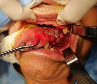
The neck physical examination reveals the presence of 2 cm x 3 cm level II and III hard, fixed, and palpable adenopathies. Right side without palpable adenopathies.
In collaboration with the clinical oncology unit of Hospital Central del Instituto de Previsión Social, an incisional biopsy was performed under local anesthesia that results in a mucosal pigmented melanoma.
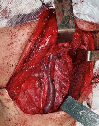
The contrast computed tomography scan performed reveals the presence of adenomegalies in the left carotid-jugular chain. In the submaxillary gland multiple adenomegalies, loss of central hilum, and conventional architecture with central necrosis are reported. The most probable diagnosis is lymphatic metastasis in one adenopathy with apparent infiltration to the internal jugular vein without cleavage plane with respect to it. No other lesions were found in different organs, thus leading to the diagnosis of primary tumor of the oral cavity.
A hemiglossectomy without reconstruction with safety margin (1.5 cm) is performed with left supraomohyoid cervical emptying followed by expectant treatment with ultrasound follow-up of the right side of the neck, and exodoncy of dental pieces 3.3 and 3.4. (See Figures 2 and 3)
The patient underwent surgery uneventfully and with good clinical progression. Aspiration drainage was, then, removed. The swallowing and talking functions of the patient were mildly impaired. The patient showed no major complications, favorable results, and was discharged 5 days after surgery.
Final pathology report: presence of a 3 cm x 3 cm x 2.5 cm mucosal pigmented melanoma with neoplasm infiltrating skeletal musculature with spread foci towards the ducts of sublingual mucosal glands without vascular or perineural invasion, and uncompromised borders. Metastatic pigmented mucosal melanoma in 3/22 nodes (macro-metastasis with capsular rupture). Cancer stage T3 N1 M0.
The patient is referred for chemotherapy with temozolomide + cisplatin with 6 cycles of local radiotherapy. Afterwards, the patient underwent follow-up every 4 months within the first year after surgery. No local relapse was ever reported. The patient was treated with phonoaudiology due to the presence of minor difficulties swallowing and talking, still with good gradual disease progression.
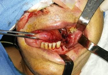
Figure 2. Result of hemiglossectomy with primary suture of the borders with polyglactin suture material 3/0. Dental pieces 3.3 and 3.4 were removed.
DISCUSSION
Due to the rarity of OMM little is known on its ethiopathogenesis and, up until today, no risk factors involved have been identified. The association between UV exposure and skin melanoma is not present in mucosal melanoma. The association between HPV, HIV and OMM has not been established either. It has been suggested that formaldehyde could be a risk factor for nasosinusal melanoma, and that oral melanoma can be preceded by a phenomenon of oral melanosis1.
Although smoking can induce the appearance of pigmented lesions in the oral mucosa, there is not enough evidence available to this date to consider tobacco as a carcinogen associated with mucosal melanoma1.
In a report from the database of the National Cancer Database of the Commission on Cancer of the American College of Surgeons including 84 836 cases of skin and non-skin melanoma from all anatomical regions, only 1.3% were cases of mucosal melanoma 55% of which were located in head and neck regions2.
The symptoms of oral mucosal melanoma include bleeding and, rarely, pain that often has a late onset. As a matter of fact, bleeding has been reported as the most common sign at diagnosis while the presence of melanic pigments has been reported in a third of the patients prior to diagnosis. Therefore, and probably associated with a later diagnosis, oral mucosal melanoma has been associated with a more sober prognosis3,5.
Tumor is often diagnosed when it causes local pain, bleeding or dental mobility. It is in these cases that the disease is in an advanced stage with presence of locoregional or distant metastases. Bleeding and color changes are often the main cause for consultation in most patients. Lymph node and distant metastases often occur in these patients being distant spread the main cause of death.
Up to this date, surgery is still the treatment of choice regarding OMM with radical tumor resection with a broad margin. The arrival of other coadjuvant therapeutic modalities like chemotherapy, radiotherapy, vaccines, and immunotherapy (interleukin-2 and interferon) have increased the overall mortality rate in up to 10%2.
Several studies claim that surgical resection with broad margins like the first-line therapy offers the best possibilities of achieving proper local control and survival. The different survival rates of patients with melanoma-both skin and mucosal melanoma-is closely associated with surgical margins2,3.
Neck dissection is indicated in patients with OMM and positive cervical adenopathies since lymph spread is associated significantly with the survival rate of this type of cancer. However, to this date, there is still controversy on cervical dissection in clinically negative necks4,5.
In both China and the United States, the neck is treated prophylactically especially when conditions so aggressive as this5.
Most studies on the management of this disease at cervical node level conclude that:
The oral cavity is a difficult anatomy regarding diagnosis and treatment.
Lymphatic metastasis has a grim prognosis.
The lack of consensus regarding neoadjuvant, immune, radio, and chemo therapies.
The lack of consensus regarding the efficacy of the sentinel node in head and neck melanoma5..
In our own opinion, the treatment of patients with intraoral mucosal melanomas with cervical dissection should become standardized5.
REFERENCES
1. Kaul S, Kumar P. Significance of early detection of oral malignant melanoma in improving prognosis. South Asian J Cancer. 2013 Oct-Dec; 2(4): 199. doi: 10.4103/2278-330X.119898 [ Links ]
2. Ballester Sánchez R, de Unamuno Bustos B, Navarro Mira M, Botella Estrada R. Actualización en melanoma mucoso. Actas Dermo-Sifiliográficas. marzo de 2015;106(2): 96-103. [ Links ]
3. Bobos M. Histopathologic classification and prognostic factors of melanoma: a 2021 update. Ital J Dermatol Venerol . 2021 Jun; 156(3): 300-321. [ Links ]
4. González García R, Naval Gías L, Martos PL, et al. Melanoma de la mucosa oral: Casos clínicos y revisión de la literatura. Med. oral patol. oral cir. bucal (Ed.impr.) may/jul 2005; 10(3): 264-271 [ Links ]
5. Pingarrón Martín L, Martín-Moro JG, Ma CY, Yu ZW, Zhang CP. Melanoma de mucosa intraoral: ¿enfermedad local o sistémica? Rev Esp Cir Oral Maxilofac. ene 2014;36(1):15-20. [ Links ]
Authors’ contributions: Dr. Marcelo Samudio had the study idea, drafted the manuscript, and was involved in the manuscript bibliographic search and review stage.
Received: August 27, 2022; Accepted: February 03, 2023
 Este es un artículo publicado en acceso abierto bajo una licencia Creative Commons
