


Servicios Personalizados
Revista

Articulo
 texto en
  Inglés (pdf)
 Articulo en XML
 Referencias del artículo
 Enviar articulo por email
Indicadores
-
 Citado por SciELO
Links relacionados
-
 Similares en
SciELO
Similares en
SciELO
Compartir

 Permalink
Cirugía paraguaya
versión On-line ISSN 2307-0420
Cir. parag. vol.46 no.3 Asunción dic. 2022
https://doi.org/10.18004/sopaci.2022.diciembre.26
Case report
Complete intestinal occlusion due to plastered Meckel’s diverticulitis

1Universidad Nacional de Asunción, Facultad de Ciencias Médicas, Hospital de Clínicas. San Lorenzo, Paraguay
La anomalía congénita más común gastrointestinal es el divertículo de Meckel, es un divertículo verdadero, ya que contiene todas las capas del intestino. Generalmente es asintomático. El diagnóstico en el 80 % restante es incidental y se hace por hallazgos quirúrgicos. Tiene un riesgo de complicación de 2-40%, siendo las más frecuentes la hemorragia, la obstrucción intestinal y la diverticulitis. La forma oclusiva de presentación es infrecuente y requiere un elevado índice de sospecha.
Palabras clave: Divertículo; Meckel; Oclusión
The most common congenital GI anomaly is Meckel's diverticulum. These are true diverticula because they contain all the layers of the intestine. It is often asymptomatic. Diagnosis in the remaining 80% of the cases is incidental and achieved through surgical findings. The rate of complications is somewhere between 2% and 40%, the most frequent of which are hemorrhage, bowel obstruction, and diverticulitis. The occlusive form of presentation is rare and requires a high index of suspicion.
Keywords: Diverticulum; Meckel; Occlusion.
Introduction
The incidence rate of Meckel’s diverticulitis in the overall population is between 1% and 2%. Clinical signs appear in 20% of the cases. Diagnosis—in the remaining 80% of the cases—is purely incidental and achieved through surgical findings. It has been reported in up to 2% of the autopsies.1,2 It is the most common GI tract disorder, and consists of the remnant of the vitelline duct proximal segment (omphalomesenteric).3 It can remain asymptomatic or present with different signs and symptoms. (4) In adults, it is often not taken into consideration regarding differential diagnosis due to the lack of suspicion and complexity regarding detection5. The most common complications are bleeding, obstruction, perforation or diverticular inflammation6. Small bowel obstruction due to Meckel’s diverticulitis has been reported in older children and adults7. Overall, this particular variety is rare. It poses a serious threat, requires a high index of suspicion, and the necessary expertise from the treating surgeon8. These elements motivate the presentation of this case report to describe the clinical presentation and surgical characteristics of mechanical bower obstruction due to Meckel’s diverticulitis in an adult.
Case report presentation
This is the case of a 30-year-old man who presents to the ER with pain of 3-day evolution followed by abdominal distension, nausea, and repeat vomiting. Twenty-four hours prior to admission he reports constipation and gases also feeling feverish twice. The patient’s only past surgical history includes one open appendicectomy 2 years ago. The abdominal physical examination reveals asymmetry due to McBurney’s point appendicectomy scar. The abdomen looks distended, painful over the right iliac fossa with defense and pain to decompression and increasing hydro-aerial noises with metal timbre. Digital rectal exam without specific findings. Lab test results reveal leukocytosis, neutrophilia, hemoglobin, and hematocrit levels of 18 000/µL, 95%, 16.4 mg/dL, and 47%, repectively. The abdominal x-ray performed confirms the presence of central dilated thin loops with multiple hydro-aerial levels. Exploratory laparotomy is indicated with diagnosis of complete bowel occlusion due to probable bands and adherences.
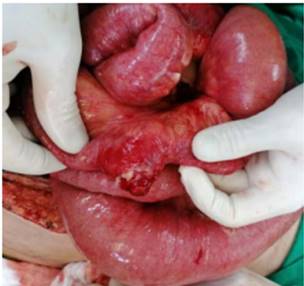
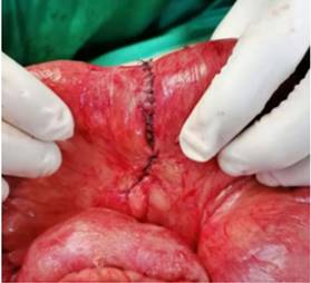
The surgery performed via supra-para-infraumbilical approach confirms the presence of plastic process with compromise of thin loops and epyplon, and adherences to the sigmoid colon. Blunt dissection maneuver is attempted, and a transition region is confirmed 140 cm away from the ileocecal valve and 180 cm from the fixed bowel loop in the thin loop antimesenteric border where there is evidence of inflamed diverticulum with necrotic core and fibrin adherences (Figure 1). Approximately 10 cm of thin loops are resected followed by manual termino-terminal anastomosis (Figure 2).
Histological study reveals the presence of a small bowel segment with wall evagination showing all components surrounded by intestinal epithelium with necrotizing acute transmural inflammatory process perforating the wall consistent with true diverticulum (acute transmural diverticulitis with peritonitis).
At the hospital ward, patient’s progression was good, and he was discharged 5 days after hospitalization with outpatient follow-up.
Discussion
Meckel’s diverticulitis appears in the antimesenteric border of distal ileum at an approximate distance of 30 cm to 60 cm from the ileocecal valve. However, cases of up to 180 cm have been reported1,2. Onset is often asymptomatic. These are some of the complications that have been reported: GI bleeding due to the presence of gastric or ectopic pancreatic tissue, bowel tumors like GI stromal tumors, bowel obstruction, perforation or diverticular inflammation3,4.
Obstructive symptomatology is the second most common way of presentation. The mechanisms of obstruction are varied like invagination, volvulus, internal hernia, diverticulitis with adherences, mesodiverticular band, foreign body impacted in the diverticulum or inclusion of the diverticulum in a true knot formed between the ileum and the sigmoid colon5. The case presented here was due to diverticulitis with plastered adherences to sigmoid colon. Diagnosis is rarely achieved in the postoperative period and can only be achieved with guarantess if the diverticulum is seen at the obstruction site6. As in the case presented here where diagnosis of bowel occlusion was achieved no Meckel’s diverticulitis was suspected as the main cause, and diagnosis was achieved during surgery. The difficulty with preoperative diagnosis is not only the result of symptoms overlapping with other conditions, but also due to the difficulty trying to identify Meckel’s diverticulitis in the imaging modalities7. Treatment should be surgical by resecting the diverticulum with a small safety margin8. In the case presented here, bowel occlusion symptoms, surgical finding of Meckel’s diverticulitis in a position further away than usual (at 140 cm from the ileocecal valve), and contact with sigmoid colon, it was deemed necessary to resect 10 cm of thin loop leaving the bowel tissue without inflammatory compromise with further ileum-ileal anastomosis.
We conclude that Meckel’s diverticulitis has very many different ways of clinical-radiographic presentation, which complicates its diagnosis. Also, we submit that bowel occlusion due to Meckel’s diverticulitis is a cause for occlusive syndrome that should be taken into consideration while performing surgery. Therefore, the general surgeon should be ready to face it with updated knowledge.
REFERENCES
1. Ruiz Borrego J. Características clínicas del divertículo de Meckel en la población infantil. Rev Gastroenterol Perú. 1995; 15(3): 247-54. [ Links ]
2. Darlington CD, Anitha GFS. Meckel's Diverticulitis Masquerading as Acute Pancreatitis: A Diagnostic Dilemma. Indian J Crit Care Med. 2017; 21(11):789-92. [ Links ]
3. Chatterjee A, Harmath C, Vendrami CL, Hammond NA, Mittal P, Salem R, et al. Reminiscing on Remnants: Imaging of Meckel Diverticulum and Its Complications in Adults. AJR Am J Roentgenol. 2017; 209(5):287-6. [ Links ]
4. Lequet J, Menahem B, Alves A, Fohlen A, Mulliri A. Meckel's diverticulum in the adult. J Visc Surg. 2017; 154(4): 253-9. Disponible en https://europepmc.org/article/med/28698005. [ Links ]
5. Aguayo P, Fraser JD, Peter SD, Ostlie DJ. Perforated Meckel`s diverticulum in a micropremature infant and review of the literature. Pediatr Surg Int. 2009; 25(6):539-41. [ Links ]
6. Cengiz F, Chabowski M, Szymanska-Chabowska A, Dorobisz T, Janczak D, Jelen M, et al. A massive bleeding from a gastrointestinal stromal tumor of a Meckel's diverticulum. Srp Arh Celok Lek. 2016; 144(3-4):219-21. [ Links ]
7. Figueredo Mari´n B, Amarilla Wildberger E, Zarza Quintana L. Divertiulitis de Meckel perforada en paciente adulto. Rev. Cir. Parag. 2018; 42(3):36-37. Doi: 10.18004/sopaci.2018.diciembre.36-37 [ Links ]
8. Miyata S, Bliss DW. A gastrointestinal stromal tumor found in perforated Meckel's diverticulum. Surg Case Rep. 2016; 2(1):67. Disponible en https://doi.org/10.1186/s40792-016-0196-8. [ Links ]
5Authors’ contributions: All the authors collaborated looking for information, collecting data, drafting the manuscript, critically reviewing it, and approving its final version.
Received: March 23, 2022; Accepted: September 19, 2022
 Este es un artículo publicado en acceso abierto bajo una licencia Creative Commons
