


Serviços Personalizados
Journal

Artigo
 texto em
  Inglês (pdf)
 Artigo em XML
 Referências do artigo
 Enviar este artigo por email
Indicadores
-
 Citado por SciELO
Links relacionados
-
 Similares em
SciELO
Similares em
SciELO
Compartilhar

 Permalink
Cirugía paraguaya
versão On-line ISSN 2307-0420
Cir. parag. vol.48 no.1 Asunción abr. 2024
https://doi.org/10.18004/sopaci.2024.abril.31
Case report
Generalized peritonitis of appendicular origin with intraoperative finding of situs inversus abdominalis

1Hospital Regional de Concepción, Servicio de Cirugía, Concepción, Paraguay
Situs inversus is a very rare congenital disease that affects one in 10,000 people. The revelation of situs inversus due to a peritoneal syndrome is an extremely rare event. Patients with situs inversus have a 0.016-0.024% incidence of acute appendicitis and generally present with some complication. We present the case of a patient with generalized peritonitis of appendiceal origin with a casual intraoperative finding of situs inversus abdominalis.
Keywords: Appendicular peritonitis; Situs inversus
El situs inversus es una enfermedad congénita muy rara que afecta a una de cada 10 000 personas. La revelación desitus inversuspor un síndrome peritoneal es un evento extremadamente raro. Los pacientes con situs inversus tienen una incidencia de 0,016-0,024 % de sufrir apendicitis aguda y generalmente se presentan con alguna complicación. Se presenta el caso de una paciente con peritonitis generalizada de origen apendicular con hallazgo intraoperatorio casual de situs inversus abdominalis.
Palabras Claves: Peritonitis Apendicular; situs inversus
INTRODUCTION
Situs inversus is a rare congenital disease that affects one in every 10,000 people. It’s characterized by specular image transposition of the abdominal and thoracic organs. The situs inversus diagnosis is generally performed incidentally while researching a non-related medical problem1.
Situs inversus’ revelation through a peritoneal syndrome is an extremely rare event. Situs inversus’ patients have a 0.016-0.024% tendency to suffer acute appendicitis and it generally presents itself with some complication. This condition can be completed when both the thoracic and abdominal organs are transposed, or partially when only one of these cavities is affected.
Acute peritonitis in situs inversus is a diagnostical dilemma due to the appendix’s abnormal position. It’s assumed that, even if the viscera is transposed, the nervous system may not show the corresponding transposition, which leads to confusions regarding vitals and symptoms. Hence, regarding the 18.4 to 31% of situs inversus’ patients, the pain caused by the left acute appendicitis has been reported on the lower right quadrant2.
In general, surgical diseases’ diagnosis in these patients is delayed due to low clinical suspiciousness, hence why usually these patients are diagnosed in later stages of the disease. In these types of patients, the diagnosis can be based on the physical exam, X-ray, and ultrasound, although CT scans have proved to have increased sensibility and specialty and has become an essential tool to handle these patients. With a male-to-female 3:2 proportion, it has an autosomal recessive presentation, but its precise genetical mechanism is yet to be identified3.
The following clinical case is presented with the objective of making the main characteristics of this unpredictable finding known in most of its presentations.
CASE PRESENTATION
43-year-old female patient reports to the emergency services with a 7-day history of evolutive abdominal pain of sudden origin to the left iliac fossa level, stinging and continuous which partially yields with common self-administered oral analgesics and covers the entire abdomen after a few days, also accompanied by vomiting on several occasions and diarrheal stools. Forty-eight hours after their admission the patient presents feces and gas detention. Physical exam yields abdominal distension, little depressiveness, very painful in all quadrants, with peritoneal defense and irritation, increased sonority, and absence of hydro-aerial sounds.
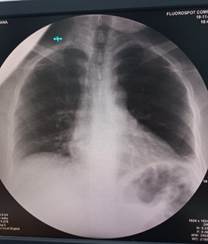
Laboratorial studies yield 10.4gr/dl hemoglobin, 29.8% hematocrit, 15,000/mm3 leukocytosis, and 88% neutrophilia. Posteroanterior thoracic and standing abdominal X-rays are requested, which yield hydro-aerial levels and great dilation of the left colonic framework. The thoracic X-rays does not yield pneumothorax, heart in its normal location. (see image 1 and 2). There are no other preoperatory image methods.
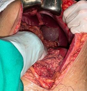
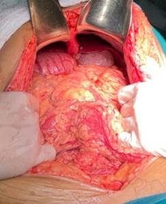
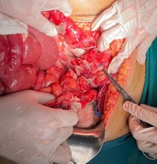
An emergency exploratory laparotomy is decided upon, with suprainfraumbilical handling, abundant fetid purulent liquid release is presented immediately after incision, approximately 700 cc suctioned, cavity exploration yields situs inversus abdominalis, liver and gallbladder occupying left hypochondrium and stomach and right-side vessel (see image 3 and 4). Colon exploration yields cecum in the left iliac fossa with cecal appendix characteristically gangrenous, punctured in the middle third and healthy base. (see image 5). Appendectomy and profuse cavity cleanse are performed.
The patient presented proper postsurgical evolution, with antibiotic treatment, being discharged after the fourth postoperative day.
Subsequent anatomopathological report shows cecal appendix with necrosis on both its distal thirds compatible with inflammatory process in all of its extension.
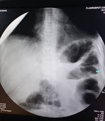
Image 1. Standing abdominal X-ray where hydro-aerial levels and colonic dilation of the left side can be observed.
DISCUSSION
Acute inflammatory process’ association with situs inversis abdominalis is extremely rare. These cases are intraoperative surprises with an 0.001% incidence in operated patients for presumed acute appendicitis. The clinical diagnosis of this type of pathology is hard to establish in absence of a previous image, especially if the symptoms are atypically located4).
Currently, the increased availability of imagery methods have proved that situs inversus are not exclusive to pediatric age and neither are always associated to comorbidity, on the contrary, they can be presented in adults and as an incidental finding5.
Even though in the presented case imagery studies such as ultrasound and CT scan were not performed, the handling and treatment is the same, hence counting on them would serve more as a presurgical guide facilitating the situs inversus’ diagnosis and diagnostical suspiciousness.
REFERENCES
1. Alonazi I, Alharthy Y, Alghamdi G. Sleeve gastrectomy in a patient with situs inversus: a case report. J Surg Case Rep. 2022 Jul 30;2022(7): rjac325. doi: 10.1093/jscr/rjac325. PMID: 35919692; PMCID: PMC9341298. [ Links ]
2. Herrera-Moncada IC, Zuluaga-Restrepo JD, Meza M, Apendicitis aguda en situs inversus totalis: reporte de un caso. Rev CES Med 2012; 26(2): 209-214 [ Links ]
3. Cissé M, Touré AO, Konaté I, Dieng M, Ka O, Touré FB, Dia A, Touré CT. Appendicular peritonitis in situs inversus totalis: a case report. J Med Case Rep. 2010 May 11;4:134. doi: 10.1186/1752-1947-4-134. PMID: 20459847; PMCID: PMC2876173. [ Links ]
4. Mihetiu AF, Bratu DG, Popescu OM, Juravle C, Dumitrean IE, Chicea R. A rare case of situs inversus totalis associated with sigmoid diverticulitis and appendicular agenesis. Embryological, clinical considerations and literature review. Rom J Morphol Embryol. 2021 Jul-Sep;62(3):861-867. doi: 10.47162/RJME.62.3.27. PMID: 35263418; PMCID: PMC9019675. [ Links ]
5. Corral G, Labra A, Schiappacasse G. Manifestaciones abdominales de las anomalías del Situs Ambiguous en el adulto: A propósito de cuatro casos. Rev. chil. radiol. [Internet]. 2013 [citado 2024 Mar 22] ; 19(1): 38-43. Disponible en: http://www.scielo.cl/scielo.php?script=sci_arttext&pid=S0717-93082013000100007&lng=es . http://dx.doi.org/10.4067/S0717-93082013000100007 [ Links ]
AUTHOR’S CONTRIBUTION: the author handled the study’s design, data collection, draft redaction and subsequent modification and approval of the manuscript.
ETHICAL CONSIDERATIONS: ethical considerations regarding the presentation of a clinical case were respected.
Received: March 29, 2023; Accepted: April 16, 2023
 Este es un artículo publicado en acceso abierto bajo una licencia Creative Commons
