


Services on Demand
Journal

Article
 text in
  English (pdf)
 Article in xml format
 Article references
 Send this article by e-mail
Indicators
-
 Cited by SciELO
Related links
-
 Similars in
SciELO
Similars in
SciELO
Share

 Permalink
Cirugía paraguaya
On-line version ISSN 2307-0420
Cir. parag. vol.47 no.2 Asunción Aug. 2023
https://doi.org/10.18004/sopaci.2023.agosto.37
Case report
Exeresis surgery on the external region of the leg - anatomical repairs
1
 http://orcid.org/0000-0003-0702-5422
1
http://orcid.org/0009-0007-3045-0247
1
http://orcid.org/0009-0002-7405-4661
1Universidad Nacional de Asunción, Facultad de Ciencias Médicas, Cátedra de Anatomía Descriptiva y Topográfica. Asunción, Paraguay
The presented case corresponds to a patient with a low-grade soft tissue tumor measuring 5 cm in the upper third of the right leg, on the lateral side. The surgical approach involved an appropriate wide resection. In relation to the study's objective of correlating the surgical approach for resecting a soft tissue tumor located on the outer side of the upper third of the leg with the anatomical compartmental and extracompartmental elements of the leg, an anatomical dissection was performed in the Descriptive Anatomy Department, focusing on the elements of the lateral compartment of the leg. During this dissection, the following structures were identified: the peroneal muscles, the lateral branch of the common peroneal nerve or external sciatic nerve, their relationships with the head of the right fibula, and their branches - the superficial and deep peroneal nerves.
El caso presentado corresponde a una paciente con un tumor de partes blandas del tercio superior de la pierna derecha, cara externa; de bajo grado de 5 cm. cuya cirugía tuvo un enfoque de resección amplia adecuada. En relación con el objetivo del trabajo de relacionar el abordaje quirúrgico de la resección de un tumor de partes blandas ubicado en la cara externa del tercio superior de la pierna con los elementos anatómicos compartimentales y extra compartimentales de la pierna, fue realizada una disección anatómica en la Cátedra de Anatomía descriptiva haciendo énfasis en los elementos del compartimiento lateral de la pierna, en donde fueron identificados: los músculos peroneos, la rama externa del nervio ciático externo o peroneo común, sus relaciones con la cabeza del peroné lado derecho y sus ramas: nervios peroneo superficial y profundo.
Palabras Clave: Disección anatómica; elementos compartimentales
Keywords: Anatomical dissection; compartmental elements
INTRODUCTION
Soft tissue sarcomas account for about 1% of all adult cancers, but this proportion increases to nearly 10% in children under 15 years old. These tumors constitute a heterogeneous group of neoplasms with still relatively unknown etiology. The overall survival of patients with soft tissue and connective tissue sarcomas is around 50% at five years after treatment initiation, with tumors located in the extremities having a more favorable prognosis than those in other locations1.
As far as surgical treatment of sarcomas is concerned, it has dramatically changed over the last three decades. Forty years ago (1970-1980), it involved radical surgery with resection of the entire muscular compartment or amputation of the limb. Nowadays, limb-sparing surgery can be performed in over 80% of cases. In oncological surgery, the objectives of surgical resection are complete removal of the tumor with wide margins and minimal functional compromise, avoiding postoperative sequelae or morphological deformities.
A proper understanding of anatomical elements allows for approaching a topographical region or performing a compartmental or extracompartmental surgery, considering anatomical relationships mainly with the vasculonervous axis, bony structures, and other adjacent tissues.
The specific objective of this study is to identify landmarks and anatomical elements of the external compartment of the upper third of the leg, both in anatomical dissection and in surgery, with an emphasis on the course of the external sciatic or common peroneal nerve, as well as its bony and muscular relationships, and the identification of its branches.
CASE PRESENTATION
A 68-year-old patient presented to a surgical service with a complaint of pain on the outer side of the upper third of the right leg, persisting for 6 months.
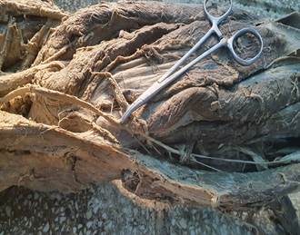
Physical examination revealed a slightly immobile soft tissue tumor. Further extension studies, including ultrasound and CT scans, were conducted, along with a percutaneous biopsy confirming the diagnosis of a soft tissue tumor on the outer side of the upper third of the right leg. A compartmental surgery was performed, preserving the common fibular nerve and using the head of the fibula as a landmark (Figures 1 and 2). The anatomopathological diagnosis reported a soft tissue tumor with clear margins. Figure 3 shows the cadaveric dissection of the common fibular nerve.
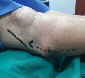
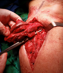
Figure 2. Common fibular nerve (external popliteal nerve), identifying the head of the right fibula.
DISCUSSION
For a low-grade tumor in a patient, surgery and proper dissection of the region are crucial, with a conservative approach to the lower limb and the identification of important vasculonervous elements in the region, considering aesthetics, gait, and patient prognosis1-2. In the specialized literature, great importance is given to compartmental and wide-margin surgery3. The treatment of a soft tissue tumor is dependent on the lesion size4-5 and anatomical compartment of the areas7-8, and the patient's follow-up through regular physical examinations and imaging studies must also be considered.
REFERENCES
1. De Vita V, Lawrence T, Rosenberg SA. Cancer: principio y practicas oncológicas. Amolca; 2017.p.1241-1250. [ Links ]
2. American Joint Committee on Cancer. AJCC Cancer staging Manual. 7ma ed. Springer; 2017. p.507-508 [ Links ]
3. Lopes A. Sarcomas de partes Moles. Rio de Janeiro Brasil:Medsi; 1999. p.129-130 [ Links ]
4. Adler RS. Musculoeskeletal interventions. En: Rumack CM, Levine D, eds. Diagnostic ultrasound. Filadelfia EUA: Elsevier; 2018. p.898-914 [ Links ]
5. Scout L, Norton ML. Ecografía general. Elsevier; 2017. [ Links ]
6. Bouchet A, Cuilleret J. Cap. 11 Región posterior de la pierna. En: Bouchet A, Cuilleret J Anatomía descriptiva, topográfica y funcional. Miembros inferiores. Ed. Med. Panamericana 1984; p.183-202 [ Links ]
7. Latarjet M, Ruiz Liard A. Anatomía Humana (4a. ed.). Buenos Aires: Médica Panamericana; 2013. [ Links ]
8. Webb WR, Brant WE, Major NM. TAC Body. Madrid: Marban; 2010. [ Links ]
7Authors Contributions: HAOF contributed to surgery and final manuscript writing; CTAL, MRGC, MMOC, GEOB contributed to anatomical dissection and image acquisition.
8Ethical considerations: This research adheres to the Nuremberg Code and the Declaration of Helsinki.
Received: January 19, 2022; Accepted: August 18, 2023
 Este es un artículo publicado en acceso abierto bajo una licencia Creative Commons
