


Services on Demand
Journal

Article
 text in
  English (pdf)
 Article in xml format
 Article references
 Send this article by e-mail
Indicators
-
 Cited by SciELO
Related links
-
 Similars in
SciELO
Similars in
SciELO
Share

 Permalink
Cirugía paraguaya
On-line version ISSN 2307-0420
Cir. parag. vol.47 no.2 Asunción Aug. 2023
https://doi.org/10.18004/sopaci.2023.agosto.9
Original article
Characterization of parotid tumor surgeries and pathological findings at the IPS Central Hospital in the period 2016-2020
1
 http://orcid.org/0000-0002-2279-5620
1
http://orcid.org/0000-0003-2733-7853
1
http://orcid.org/0000-0002-7319-7479
1Hospital Central del Instituto de Previsión Social. Asunción, Paraguay.
Introduction:
Parotid tumors are considered a relatively rare pathology, comprising a group of tumors that affect the head and neck. Within salivary gland pathology, they constitute 80%, and 3% of all head and neck tumors. Of these, 80% are benign tumors. The objective is to characterize parotid tumor surgeries and anatomopathological findings at the Central Hospital of IPS during the period 2016-2020.
Methodology:
Observational, descriptive, and retrospective study of the database, with clinical records of patients with a history of parotid tumor surgery at the Central Hospital of IPS during the period 2016-2020.
Results:
A total of 44 parotid surgeries were analyzed. There was no gender predominance. Patients aged 51 to 60 were the most affected. Pleomorphic adenoma was the most common benign tumor, followed by Warthin's tumor. Among malignant tumors, various types of carcinomas accounted for 9% of cases. Superficial parotidectomy was performed in 64% of cases.
Conclusion:
The majority of parotid tumors were benign, with pleomorphic adenoma being the most common. Superficial parotidectomy was performed in nearly 2/3 of the cases.
Keywords: parotid tumor; parotidectomy; general surgery
Introducción:
Los tumores parotídeos son considerados una patología relativamente rara, constituyendo un grupo de tumores que afectan a la cabeza y el cuello. Dentro de la patología tumorales de las glándulas salivales, constituyen un 80%, y el 3% de todos los tumores de cabeza y cuello. De estos, un 80% corresponde a tumores benignos. Como objetivo es caracterizar las cirugías de tumor de parótida y hallazgos anatomopatólogicos en el Hospital Central del IPS en el periodo 2016-2020.
Metodología:
Estudio observacional, descriptivo y retrospectivo de la base de datos, con fichas clínicas de pacientes con antecedente de cirugía por tumor de parótida en el Hospital Central del IPS en el periodo 2016-2020.
Resultados:
Fueron analizadas un total de 44 cirugías de parótida. No hubo predominio de sexo. Los pacientes de 51 a 60 años fueron los más afectados. El adenoma pleomórfico fue el tumor benigno más frecuente, seguido del tumor de Warthin. Entre los tumores malignos, distintos tipos de carcinomas representaron el 9% de los casos. La parotidectomía superficial fue realizada en 64%.
Conclusión:
La mayoría de los tumores de parótida fueron benignos, con el adenoma pleomórfico como el más frecuente. En casi 2/3 de los casos se realizó parotidectomía superficial.
Palabras claves: tumor de parótida; parotidectomía; cirugía general
INTRODUCTION
Parotid tumors are considered a relatively rare pathology, comprising a diverse group of tumors that affect the head and neck. Within salivary gland pathology, they constitute 80%, and 3% of all head and neck tumors. Of these, 80% are benign tumors1-3.
These tumors are characterized by a wide diversity in their pathological characteristics; the pathological classification of these tumors by the World Health Organization describes nearly 10 different forms of adenomas, with pleomorphic adenoma and Warthin's tumor predominating, but also around twenty types of carcinomas, non-epithelial tumors, lymphomas, and secondary tumors. The clinical presentation varies depending on the type of tumor, particularly based on its benign or malignant nature4,5.
Pleomorphic adenoma is the most common benign tumor of the salivary glands, accounting for approximately 60% of all salivary neoplasms. It is mainly composed of a proliferation of myoepithelial cells and a wide range of components of epithelial and mesenchymal tissue, surrounded by a clear fibrous capsule. Around 80% of pleomorphic adenomas occur in the parotid gland, 10% in the submandibular gland, and 10% in the minor salivary glands of the oral cavity. The average age of presentation is 46 years, with a range spanning from the third to fifth decades of life. However, it has been found in individuals of all ages, with a slight predilection for females6,7.
Warthin's tumor is the second most common benign neoplasm of the salivary glands. It is mainly located in the parotid gland, accounting for 6-10% of all parotid tumors. It occurs in older men, but the incidence in women has been increasing, possibly related to increased smoking in this group3). As for oncocytoma, it is nearly exclusive to the parotid gland and represents less than 1% of all parotid tumors. Malignant degeneration is a rare possibility, possibly explained by an acquired genetic defect causing mitochondrial dysfunction3,8.
Mucoepidermoid carcinoma is the most common malignant neoplasm in both major and minor salivary glands, accounting for almost 30% of all malignant salivary gland neoplasms9,10). Approximately half of mucoepidermoid carcinomas occur in the major salivary glands, with 80% in the parotid gland, 8 to 13% in the submandibular gland, and 2 to 4% in the sublingual gland. The time of presentation occurs between the second and eighth decades of life, and it is the most frequent malignant tumor in individuals under 20 years of age, with a predilection for the hard palate. There is also a clear preference for the white race9.
Malignant transformation of a mixed tumor occurs about 10 to 20 years later than the benign form and is suspected based on signs and symptoms suggestive of malignancy, as previously mentioned (facial paralysis, rapid growth, pain, adherence to planes, etc.).
Adenocarcinoma accounts for about 4% of tumors. Overall, they have a strong tendency to metastasize (50% lymphatic and 30% hematogenous). There are three fundamental types: mucinous (lower survival compared to other subtypes: 30% at 20 years), salivary ductal (elderly males), and intercalated ductal (elderly, high 10-year survival).
Metastases to salivary glands can occur via lymphatic, hematogenous, or contiguous spread (more common in soft tissue sarcomas, bone tumors, and skin tumors). The most common histological types of lymphatic spread are cutaneous squamous cell carcinoma and melanoma, while the most common origin in hematogenous spread is the lung2.
Parotid tumors typically present as progressive volume enlargement localized to this region. Facial motility alteration due to facial nerve involvement, regional pain, and/or regional lymphadenopathy are suggestive findings of malignancy. Currently, in head and neck oncology, the diagnosis and treatment of these tumors remain a challenge7.
The main complementary investigations include magnetic resonance imaging and fine-needle aspiration, often guided by ultrasound. Computed tomography does not play a prominent role in the evaluation of these parotid tumors. The treatment of benign parotid gland tumors is based on surgery, with the type of resection being debated in the literature and varying between wide excision techniques, such as total parotidectomy with preservation of the facial nerve, and more or less limited procedures, such as extracapsular dissection. The treatment of malignant tumors is based on surgery and radiation therapy, with indications being based on tumor stage4.
This study aims to characterize the diagnoses and surgeries performed in patients with parotid tumors at the Central Hospital of the Instituto de Previsión Social (HC-IPS).
MATERIALS AND METHODS
The study conducted is observational, descriptive, and retrospective in nature, involving patients with a history of parotid tumor surgery at the HC-IPS in the period 2016-2020.
During data collection, operative records and biopsy reports of the studied patients were used. Patients whose operative records were incomplete or whose pathology results were unavailable in the computerized system were excluded. The database was stored from January 2016 to December 2020 in an Excel file, considering following variables: age, gender, operative findings, surgical site, procedure performed, and anatomopathological diagnosis.
After obtaining approval for the research protocol and authorization to access the Hospital Information System (SIH) database, the data were stored in an Excel template file from January 2016 to December 2020, following the inclusion and exclusion criteria mentioned above. Descriptive statistics were employed, including summary measures according to distribution, frequency table, and pie and bar charts. The data were collected, described, and tabulated in a Microsoft Office Excel spreadsheet.
RESULTS
A total of 44 parotid surgeries were analyzed during the period 2016-2020. The variables used for the study included age, sex, type of surgery performed, side, and histopathological diagnosis. The distribution according to sex was 50% for both female and male patients, respectively.
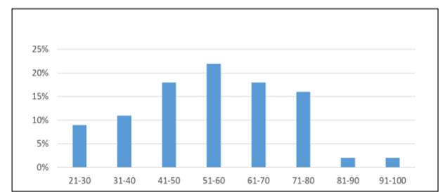
In terms of the age group of the operated patients, 22% corresponded to patients aged 51-60 years, 18% to those aged 41-50 years, 18% to those aged 61-70 years, 16% to those aged 71-80 years, 11% to those aged 31-40 years, 9% to those aged 21-30 years, 2% to those aged 81-90 years, and 2% to those over 90 years. (Graph 1)
Regarding the anatomical location, the left side was predominant over the right side, accounting for 52% and 48% respectively.
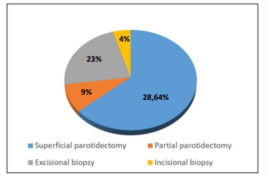
In terms of the type of operation performed, 64% underwent superficial parotidectomy, 22% excisional biopsy, 9% partial parotidectomy, and 4.5% underwent incisional biopsy. (Graph 2)
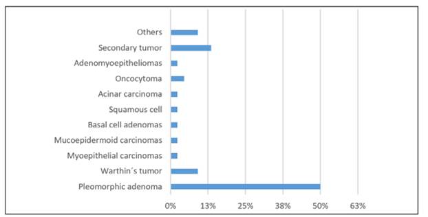
Regarding the histopathological findings, the majority were pleomorphic adenomas (50%), followed by Warthin’s tumors (9%), oncocytomas (4.54%), myoepithelial carcinomas (2.27%), mucoepidermoid carcinomas (2.27%), basal cell adenomas (2.27%), squamous cell carcinomas (2.27%), acinar cell carcinomas (2.27%), adenomyoepitheliomas (2.27%), in 13.6% of the analyzed samples, secondary tumors were found as a result of other primary entities, such as squamous cell carcinomas of the skin, and in 9.1% of cases, other types of findings were present (lipomas, lymphoepithelial cysts) (Graph 3).
DISCUSSION
There is a wide variety of diseases related to head and neck tumors, among them, tumors of the parotid gland constitute the majority of this group. Approximately 80% of parotid gland tumors are benign13. Among the most frequent are pleomorphic adenomas and Warthin's tumors, accounting for 50% and 10% of all parotid tumors, respectively. In our series, the incidence of benign tumors was 79.4% (27 cases), including only primary parotid tumors. Of these, 64% (22 cases) were pleomorphic adenomas, 11.7% (4 cases) were Warthin's tumors, and 2.9% (one case) were basal cell adenomas, which aligns with reports in the literature1,11-13. Primary malignant tumors of the parotid accounted for 20% (7 cases) in this series, slightly lower than reported in the literature. This lower rate could be due to the exclusion of tumors secondary to other organs, which accounted for 13% (6 cases) of the studied specimens.
In the international literature, Lin et al1). described a similar percentage to the one mentioned above, with a benignity rate of 85%, with the most common finding being pleomorphic adenomas in 51% of the examined samples. Regarding malignant tumors, mucoepidermoid carcinoma was the most common with an incidence of 3%.
Tapia C et al6). found results with similar characteristics. Out of 70 parotid tumors, 84.3% were benign tumors and 15.7% were malignant tumors. Among benign tumors, 76.3% were pleomorphic adenomas and 6.7% were Warthin's tumors. Among other malignant tumors, some corresponded to myoepithelial carcinoma, mucoepidermoid carcinoma, adenoid cystic carcinoma, acinar cell carcinomas, and non-Hodgkin lymphoma.
It's important to consider sociodemographic factors, as concluded by Rocha R et al12., who described that more than half of the cases are diagnosed at advanced stages due to the rural origin of a large part of the affected population, and in which the age group 45-64 (53.8%) predominated. These individuals belong to the economically and socially more active age group. They also found that the most accepted etiological factor was occupational risks and exposure to radioactive sources.
Regarding the age of presentation, what is reported in the literature is consistent with our series, both for male and female patients5,11. The incidence in women was higher for both pleomorphic adenomas and Warthin's tumors. Seven male patients presented malignant tumors, and four female patients presented malignant tumors. It's noteworthy that the diagnosis of mucoepidermoid carcinoma was made in a male patient and lymphoma in a female patient.
CONCLUSION
Parotid tumors constitute a group with diverse etiology, with a high proportion of benign pathology. No gender difference was found in the incidence, and the age range with the highest occurrence was the sixth decade of life.
The most performed surgery was superficial parotidectomy, and the most frequent pathological finding was pleomorphic adenoma.
REFERENCES
1. Lin CC, Tsai MH, Huang CC, Hua CH, Tseng HC, Huang ST. Parotid tumors: a 10-year experience. Am J Otolaryngol. abril de 2008;29(2):94-100. [ Links ]
2. Rey Biel J, Sánchez Aniceto G, Salmerón Escobar JI, Martorell Martínez V. Cap. 50. Tumores de la glándula parótida. En: Sociedad Española de Cirugía Oral y maxilofacial y de Cabeza y cuello. Ed. Protocolos clínicos de la SECOM-CyC. España: internet; 2006. p. 693-708 [ Links ]
3. Cisternas Bittencourt MJ, Corrochano EO, Saa Álvarez MR. Cap. 148. Patología tumoral de las glándulas salivales. En: Sociedad Española de Otorrinolaringología, ed. Libro virtual de formación en otorrinolaringología SEORL. Madrid, España: internet; 2015. p.1-16 [ Links ]
4. Bonfils P, Laccourreye O, Giraud P, Halimi P. Tumores de la glándula parótida. EMC - Otorrinolaringología abr 2017;46(2):1-17 [ Links ]
5. Campolo González A, Ramírez Skinner H, Vargas Díaz A, León Ramírez A, Goñi Espildora I, Solar González A. Perfil epidemiológico de neoplasias epiteliales de glándulas salivales. Rev méd Chile 2018;146(10):1159-66. [ Links ]
6. Vidal GM, Galindo PIM, Rodríguez GL, Fernández GS. Adenoma pleomorfo de la glándula submandibular. Reporte de un caso y revisión de la literatura. Revista ADM. 2019;76(6):336-342. [ Links ]
7. Neville BW, Damm DD, Allen CM, Chi AC. Oral and Maxillofacial Pathology. 4ta ed. Missouri: WB Saunders, Elsevier; 2016 [ Links ]
8. Avila RE, Samar ME, Fonseca IB, Corball AG, Carriel V, García-Martinez L, et al. Proliferaciones Oncocíticas de Glándulas Salivales: Estudio Estructural e Inmunohistoquímico de 7 Casos. International journal of odontostomatology. mar 2019;13(1):82-8. [ Links ]
9. Guevara-Canales JO, Morales-Vadillo R, Guzmán-Arias G, Cava-Vergiú CE, Guerra-Miller H, Montes-Gil JE. Mucoepidermoid carcinoma of the salivary glands. A retrospective study of 51 cases and review of the literature. Acta Odontol Latinoam. 2016 ; 29: 9. [ Links ]
10. Diwakar JK, Agarwal A, Garg C, Giri KY, Dandriyal R, Kumar G. A Rare Case of Mucoepidermoid Carcinoma of Parotid with Mandibular Metastasis. Ann Maxillofac Surg. 2019;9(1):205-7. [ Links ]
11. Tapia C M, Hernández G T, Fredes C F, Urra B A, Compan J Á, Ortega R P. Tumores de glándula parótida: Experiencia quirúrgica Hospital Guillermo Grant Benavente. Rev Otorrinolaringol Cir Cabeza Cuello. dic 2018;78(4):385-91. [ Links ]
12. Rocha Remón P, Coca Granado RM, Fonseca Pisch AJ, Rocha Remón P, Coca Granado RM, Fonseca Pisch AJ. Caracterización epidemiológica y clínico-terapéutica de las neoplasias malignas de glándulas salivales. Revista Cubana de Cirugía [Internet]. junio de 2019 [citado 1 de julio de 2021];58(2). Disponible en: Disponible en: http://scielo.sld.cu/scielo.php?script=sci_abstract&pid=S0034-74932019000200002&lng=es&nrm=iso&tlng=es [ Links ]
13. Kamal SA, Othman EO. Diagnosis and treatment of parotid tumours. The Journal of Laryngology & Otology abr 1997;111(4):316-21. [ Links ]
6Author Contributions: Dr. Pablo Schaerer, Dr. Adriana Echeverría, Dr. Ariel Benegas, Dr. Mirna Gamarra, and Dr. Martin Matoza conceived the idea, conducted the work, performed the literature search, and reviewed the final draft.
Received: November 28, 2022; Accepted: June 13, 2023
 Este es un artículo publicado en acceso abierto bajo una licencia Creative Commons
